
|
Fig. S4
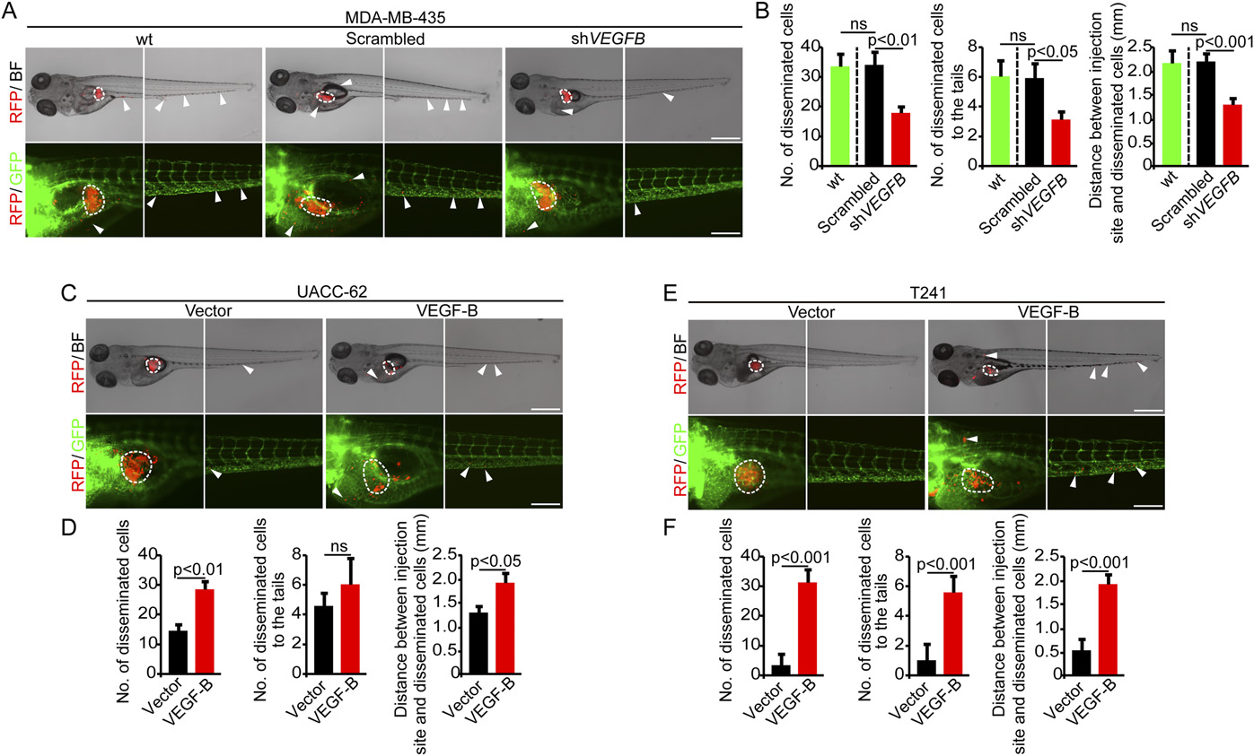
VEGF-B promotes cancer metastasis in a zebrafish model. (A) Dissemination of DiI-labeled human nontransfected WT, scrambled-shRNA-, and VEGFB-shRNA- MDA-MB-435 melanoma cells in zebrafish. Dashed lines encircle primary tumor sites. Arrowheads indicate the disseminated tumor cells. [Scale bar, 500 μm (Upper); 200 μm (Lower).] (B) Quantification of disseminated WT, scrambled-shRNA-, and VEGFB-shRNA-MDA-MB-435 melanoma cells in the whole body of zebrafish embryos (n = 10 zebrafish embryos per group). (C) Dissemination of DiI-labeled human vector- and VEGF-B-UACC-62 melanoma cells in zebrafish. Dashed lines encircle primary tumor sites. Arrowheads indicate the disseminated tumor cells. [Scale bar, 500 μm (Upper); 200 μm (Lower).] (D) Quantification of disseminated vector- and VEGF-B-UACC-62 melanoma cells in the whole body of zebrafish embryos (n = 10 zebrafish embryos per group). (E) Dissemination of DiI-labeled vector- and VEGF-B-T241 tumor cells in zebrafish. Dashed lines encircle primary tumor sites. Arrowheads indicate the disseminated tumor cells. [Scale bar, 500 μm (Upper); 200 μm (Lower).] (F) Quantification of disseminated vector- and VEGF-B-T241 tumor cells in the whole body of zebrafish embryos (n = 10 zebrafish embryos per group). All error bars represent SEM. All P values were analyzed according to Student’s t test. ns, not significant.