
|
Fig. S4
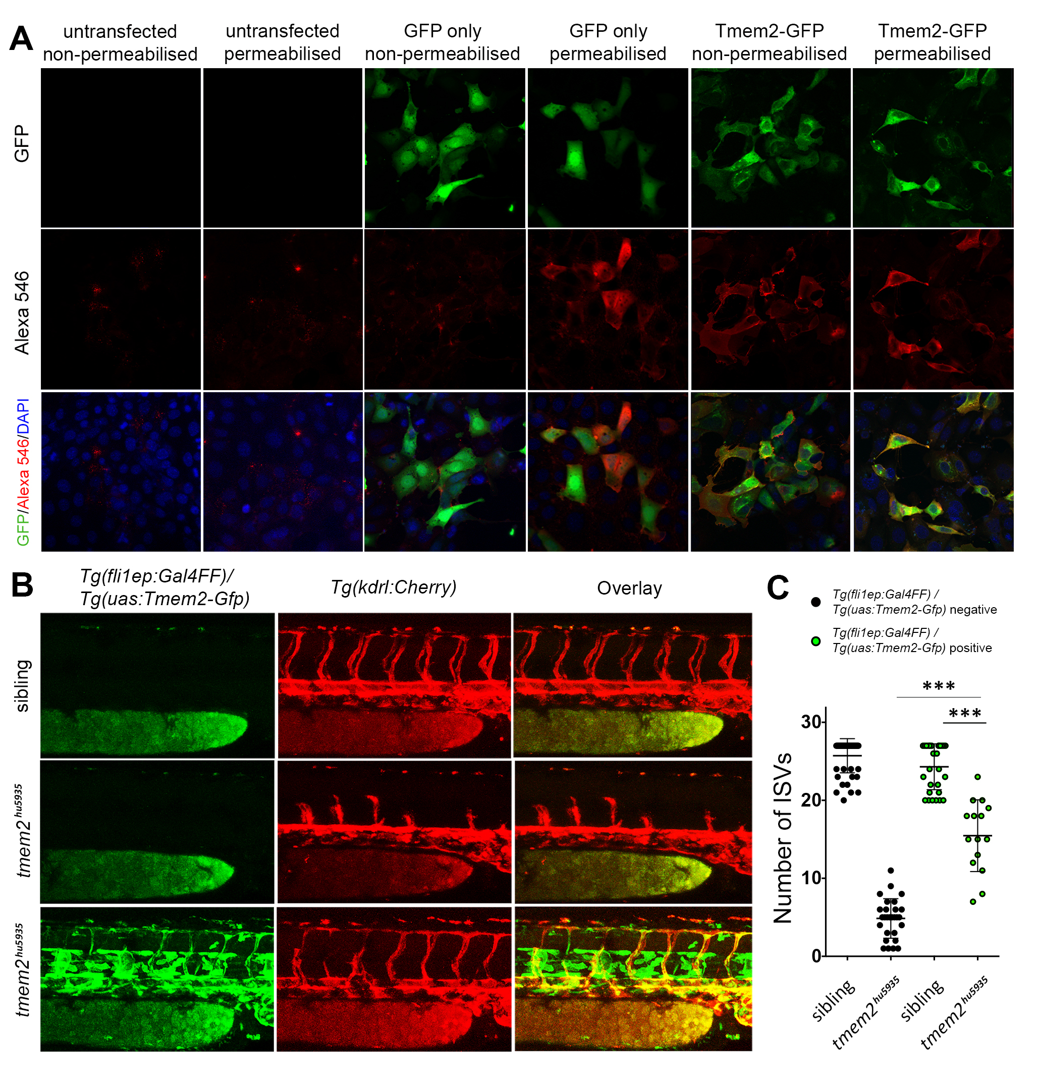
Related to Figure 2: The C-terminus of Tmem2 is localized extracellularly and Tmem2 can function in endothelial cells autonomously.
A. Permeabilisation experiment showing that mammalian cells transfected with GFP alone require permeabilisation to detect GFP using anti-GFP red immunofluorescence. Human Tmem2-GFP, by contrast, does not require permeabilisation for GFP detection, indicating that the C-terminal GFP tag localizes extracellularly. B. tmem2hu5935 mutant embryos had significantly more ISVs when overexpressing Tmem2-Gfp in the endothelium using the Tg(fli1ep:Gal4) / Tg (uas:Tmem2-Gfp) lines crossed onto the Tg(kdrl:mCherry) background, showing that Tmem2-Gfp expressed from a local endothelial source was sufficient to partially rescue angiogenesis. C. Quantification of the ISV phenotype in sibling (n=30 with; n=30 without) and tmem2hu5935 mutants with (n=14) and without (n=28) the presence of fli1ep-driven Tmem2-Gfp expression. ∗∗∗p < 0.0001; Error bars represent ±SD.
Reprinted from Developmental Cell, 40, De Angelis, J.E., Lagendijk, A.K., Chen, H., Tromp, A., Bower, N.I., Tunny, K.A., Brooks, A.J., Bakkers, J., Francois, M., Yap, A.S., Simons, C., Wicking, C., Hogan, B.M., Smith, K.A., Tmem2 Regulates Embryonic Vegf Signaling by Controlling Hyaluronic Acid Turnover, 123-136, Copyright (2017) with permission from Elsevier. Full text @ Dev. Cell