
|
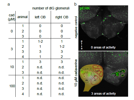
Fig. S2
Pronounced left-right symmetry of activated glomeruli
In whole mount experiments we compared number and position of pERK-labelled glomerular-shaped areas in the left and right olfactory bulb. a) Table with numbers of activated glomeruli. In all cases, where delineation of glomeruli was unambiguously possible (all experiments with 3 μM, and one with 10 μM cadaverine), we observed a (nearly) equal number of glomeruli labelled on each side. b) Images show pERKlabelling in negative control (top panel) and after exposure to 10 μM cadaverine (bottom panel). Olfactory bulbs are shown from the dorsal view. Activated glomeruli are outlined by dashed circles both in left and right olfactory bulb. A schematic glomerular map (drawn using main positional information from4) was made partially transparent and placed over the left olfactory bulb to visualize the position of cadaverine-responsive glomeruli in the dorsolateral cluster. n.d., not determined: an unambiguous identification of single glomeruli was impossible due to high signal density in the whole mount preparations.