
|
Fig. S1
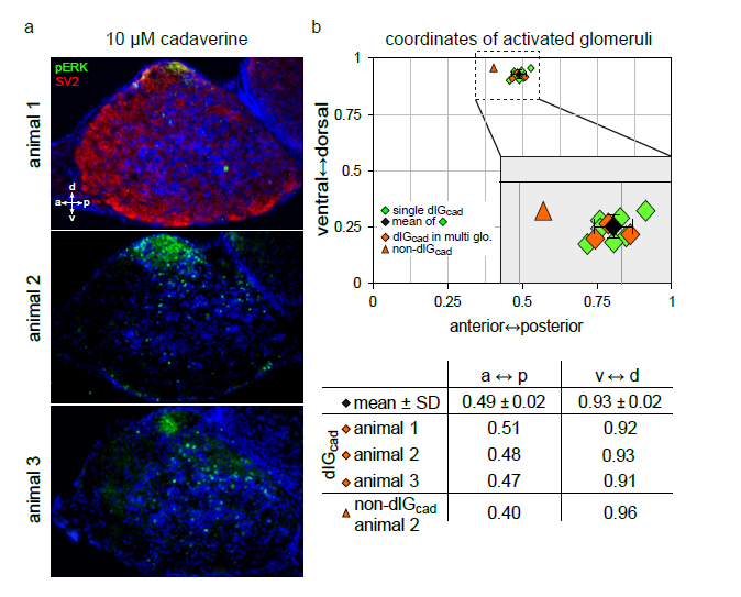
The dlGcad can also be found in bulbi with multiple pERK-labelled glomeruli
a) α-pERK labels active glomeruli in cryostat sections of the olfactory bulb from 3 different animals stimulated with 10 μM cadaverine. b) Top panel, mapped glomerular positions are shown in the coordinate system used. Green squares, glomerular position, when only a single glomerulus was labelled (n=7, from 3 animals exposed to 3 μM and 1 animal exposed to 10 μM cadaverine, respectively); black square and error bars, average position ± SD; orange squares, three examples for dlGcad positions in cases where multiple glomeruli were labelled; orange triangle, an adjacent non-dlGcad glomerulus. Note the unambiguous identification of the dlGcad glomerulus in all three cases with multiple labelled glomeruli, and the clear distinction even to the directly adjacent non-dlGcad glomerulus (this was the only case in which a non-dlGcad glomerulus was situated in the same section as the dlGcad glomerulus). Bottom panel, numeric values for coordinates of dlGcad and non-dlGcad glomeruli in experiments with multiple glomeruli labelled (orange symbols). For comparison the mean value for the 7 cases, in which a single glomerulus was labelled, is shown (black square). Note the clear distinction between positions of dlGcad and non-dlGcad glomeruli.