
|
Fig. S1
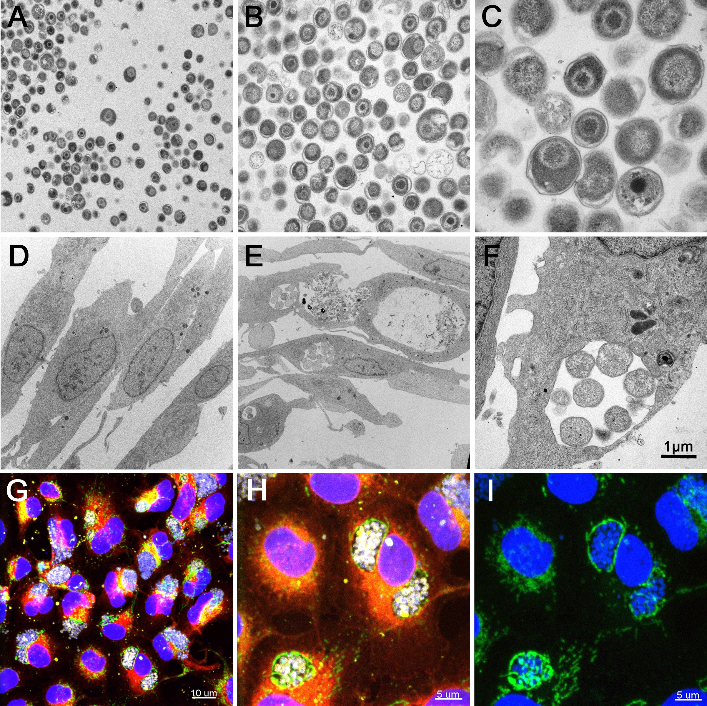
Infection of EPC cells with W. chondrophila. (A-C) TEM of W. chondrophila EBs, freshly harvested from an amoeba co-culture and injected into eggwhite. (D) TEM of non-infected EPC cells. (E) TEM of EPC cells, infected with W. chondrophila. (F) TEM of a W. chondrophila inclusion, containing replicating RBs and surrounded by host cell mitochondria. (G-I) IF staining of infected EPC cells with concanavalin A (red), an anti-Waddlia antibody (yellow), an anti-OxPhosIV antibody to stain mitochondria (green) and DAPI. The perinuclear BCVs show close association with host cell mitochondria. (I) DAPI and anti-OxPhosIV antibody alone.