
|
Fig. 6
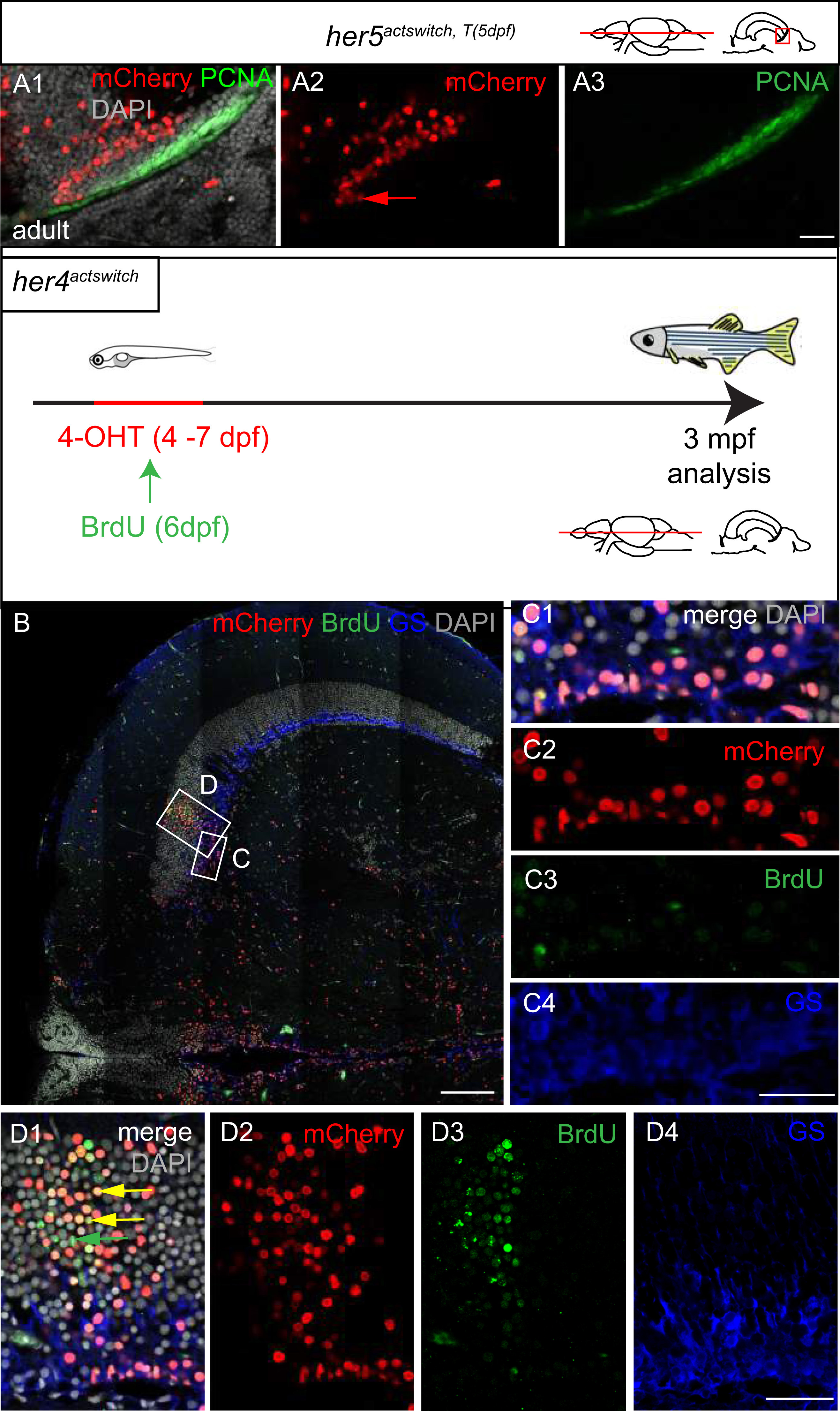
Evidence for a minor direct route of TeO neurons generation independent of her4-positive RG progenitors. A. Organization of polyclones at the TPZ border in her4actswith, T(5dpf) adults, co-immunostained for mCherry and PCNA and counterstained with DAPI. High magnification of a horizontal section, confocal microscopy. A1: merge; A2, 3: single channels. Note that polyclones extending across the entire PGZ are attached to the most ventricular domain of the TPZ. B, C. Compared fate of her4-positive and BrdU-labelled progenitors in the adult TeO. Top panel: experimental scheme. B. Low magnification of a horizontal section showing an entire TeO hemisphere in a double labelled adult, co-immunostained for mCherry, BrdU and GS, and counterstained with DAPI. C. High magnification views of the area boxed in B, located anterior to the labelled stripe. C1: merge, C1-C3: single channels. Confocal views. Note that recombination of the RG layer anterior to the stripe is complete. D. High magnification views of the stripe area, as boxed in B. D1: merge, D2-D4: single channels. Confocal views. Yellow arrows point to double mCherry and BrdU-positive neurons, green arrow to neurons positive for BrdU only. Scale bars: A, B 100 µm, C, D 50 µm.
Reprinted from Developmental Biology, 420(1), Galant, S., Furlan, G., Coolen, M., Dirian, L., Foucher, I., Bally-Cuif, L., Embryonic origin and lineage hierarchies of the neural progenitor subtypes building the zebrafish adult midbrain, 120-135, Copyright (2016) with permission from Elsevier. Full text @ Dev. Biol.