
|
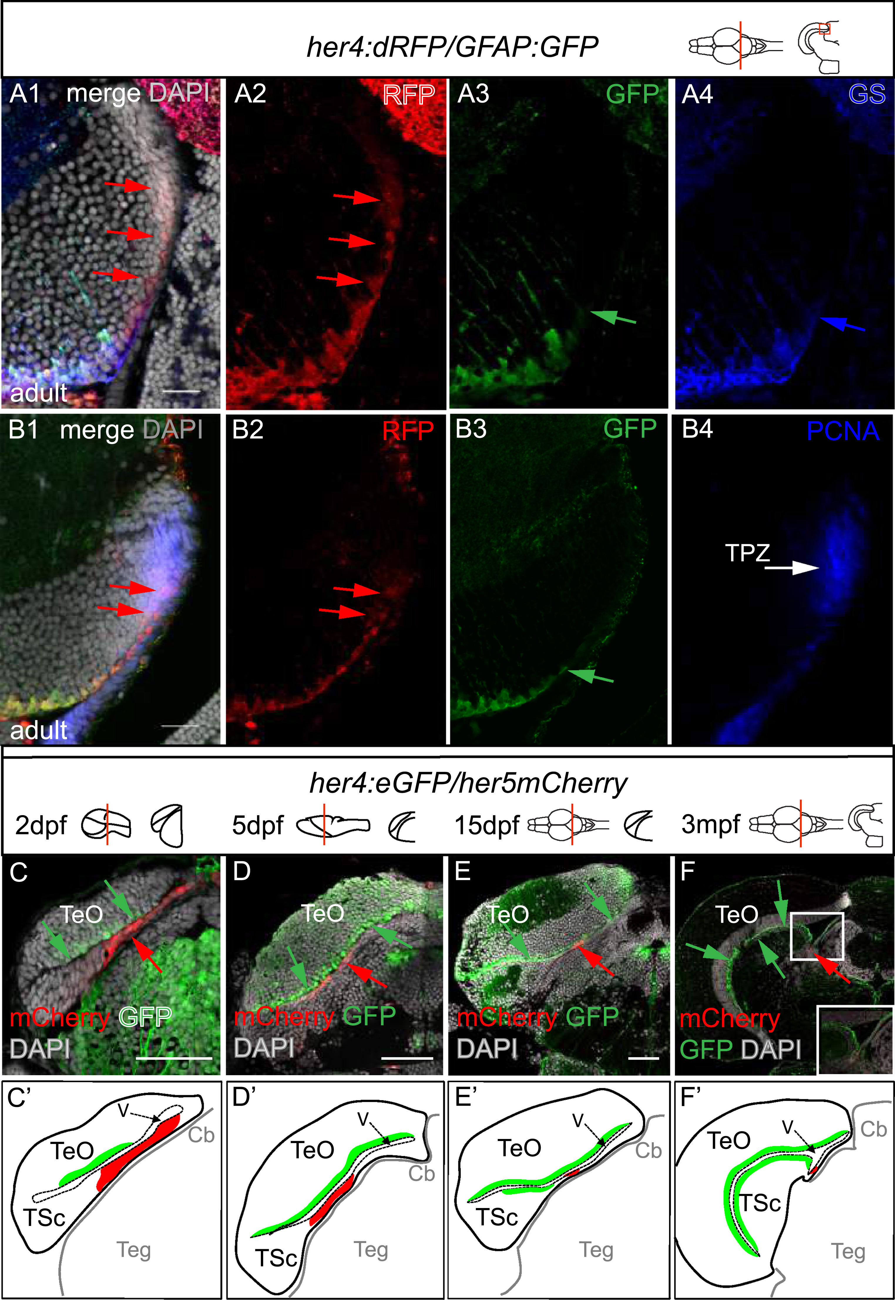
Fig. 2
The her5-positive, TPZ, her4-positive and mature radial glia domains are spatially ordered in the posterior TeO. All views are confocal images. A, B. Compared expression of Her4-RFP, GFAP-GFP and GS (A) or PCNA (B) on cross-sections of the adult midbrain in double transgenic animals (high magnifications of the area boxed in the schematic). Panels 1 are RFP, GFP and GS merge views with DAPI counterstaining, Panels 2–4 are single channel views (color-coded). Note that Her4-RFP-positive glial cells (red arrows) extend into a more lateral ventricular domain than mature RG (A 1–4, B3, green or blue arrows point to the lateral limits of GFAP-GFP and GS expression, respectively) and reach into the PCNA-positive domain (B4, white arrow). C-F. Compared position of the Her5-mCherry (red, red arrows) and Her4-GFP (green, green arrows) positive territories in the posterior midbrain at increasing stages from embryo to adult, observed on cross sections (levels and stages indicated on the schematics). C′-F′. Interpretative drawings of C-F highlighting in red the her5-positive zone and in green the area covered by the cell bodies of her4-positive RG. Scale bars: A,B 100 µm, C-F 50 µm. Abbreviations: Cb: cerebellum, TeO: tectum opticum, TPZ: tectal proliferation zone, TSc: torus semi-circularis, V: ventricle.
Reprinted from Developmental Biology, 420(1), Galant, S., Furlan, G., Coolen, M., Dirian, L., Foucher, I., Bally-Cuif, L., Embryonic origin and lineage hierarchies of the neural progenitor subtypes building the zebrafish adult midbrain, 120-135, Copyright (2016) with permission from Elsevier. Full text @ Dev. Biol.