
Image
|
Figure Caption
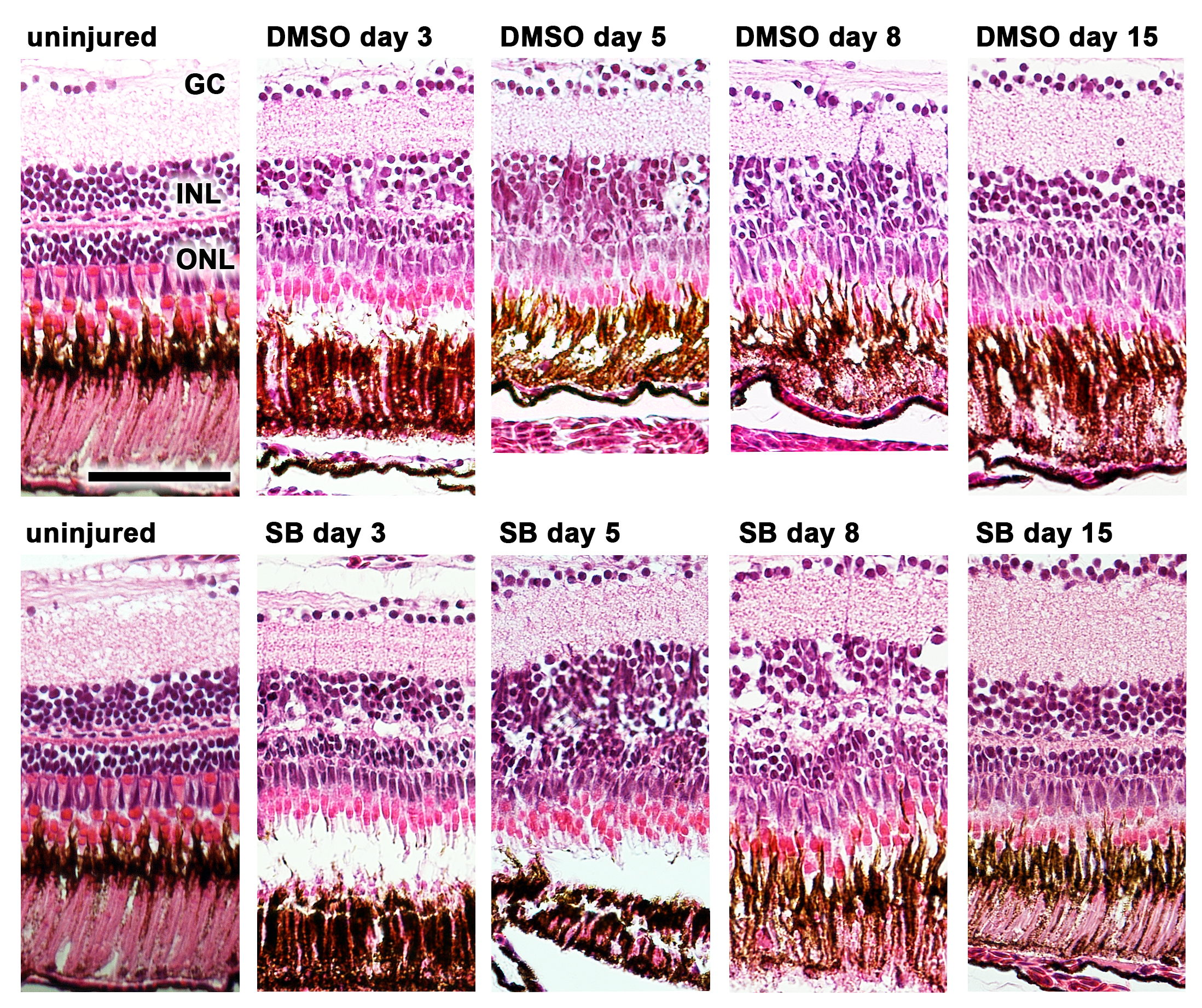
Fig. 4
H&E staining of zebrafish retinas before (uninjured) and after induction of retina degeneration with MNU.
In the non-inhibited (0.1% dimethyl sulfide, DMSO) and inhibited group (small molecule inhibitor SB431542), a reduction of rod cells was observed starting at day 3. In the non-inhibited group the reduction of rod photoreceptors persisted until day 8, whereas in the group with the inhibited TGFβ pathway (small molecule inhibitor SB431542) a rapid recovery was observed already at day 5. Scale bar indicates 50 μm. GC: ganglion cells, INL: inner nuclear layer, ONL: outer nuclear layer, SB: SB431542
Figure Data
Acknowledgments
This image is the copyrighted work of the attributed author or publisher, and
ZFIN has permission only to display this image to its users.
Additional permissions should be obtained from the applicable author or publisher of the image.
Full text @ PLoS One