
|
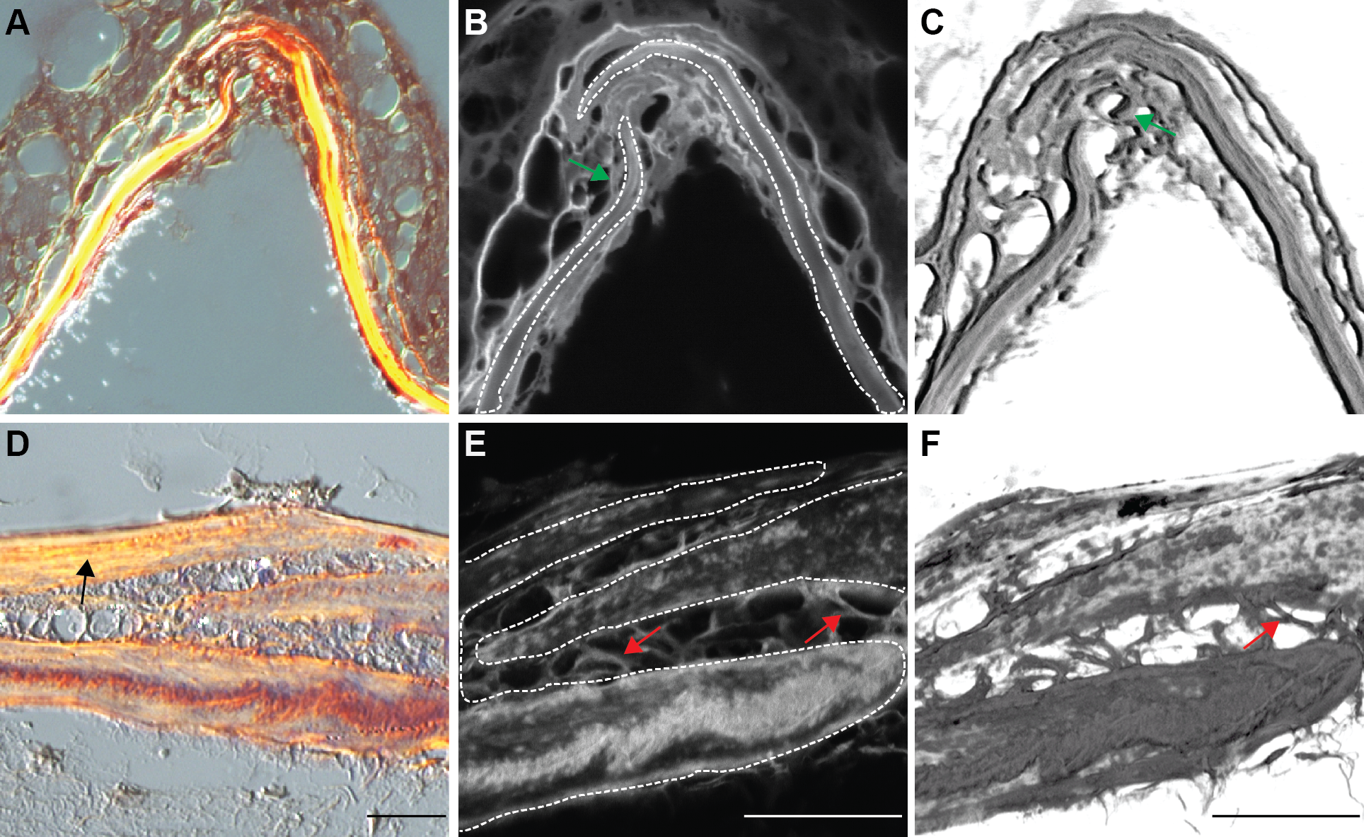
Fig. 9
Picro sirius red staining of transverse sections of the skulls.
Plane polarized light microscopy for (A, B, C) juvenile fish (13.4 mm SL) and (D, E, F) young adult fish at age of 12 wpf respectively, at 40x magnification. (B, E) confocal microscopy of the same sections at 100x magnification, and (C, F) 3D rendered confocal images at 100x magnification. The longitudinal organization of collagen fibrils is indicated by a black arrow (D). The frontal bones are outlined by white dashed lines (B, E). The left frontal plate developed a split end and the right frontal bone grew in between. The red arrow indicates fibers connecting two frontal bones; the green arrow indicates fibers that grown in between the cells. Scale bars are 20 μm.