
|
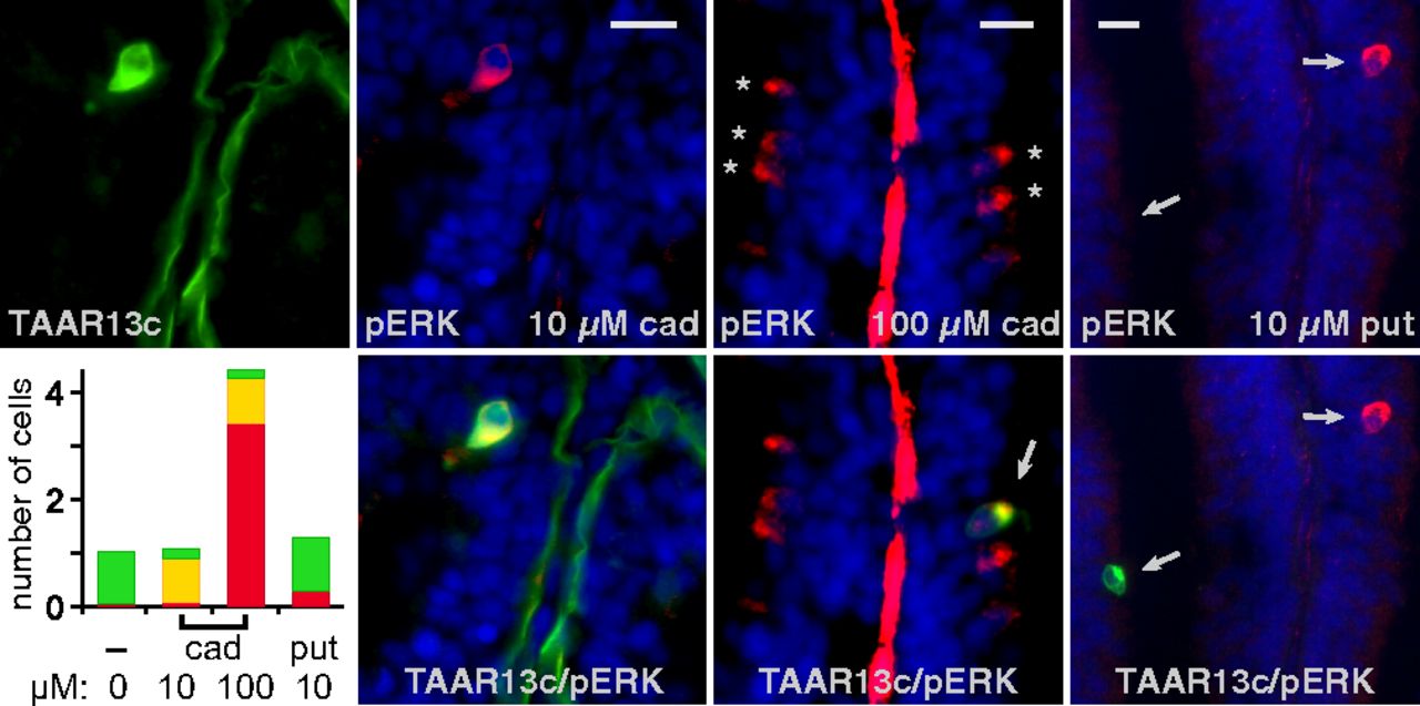
Fig. 5
Cadaverine activates TAAR13c-expressing neurons. Zebrafish were exposed to cadaverine (cad) and putrescine (put) at concentrations indicated and processed for concomitant IHC of TAAR13c (green fluorescence) and pERK (red fluorescence). DAPI was used as nuclear counterstain (blue). Sometimes the basal lamina was stained unspecifically (green and red stripes in the center of some lamellae). Asterisks, pERK+ cells; arrows, colabeled cells (yellow) and pERK+/TAAR13c− cells (red). (Scale bars: 10 μm.) (Lower Left) Quantitative evaluation, values are given as normalized cell numbers (120–250 cells per condition were analyzed); green bar, TAAR13c+/pERK− cells; yellow bar, double label; red bar, TAAR13c−/pERK+ cells.