
|
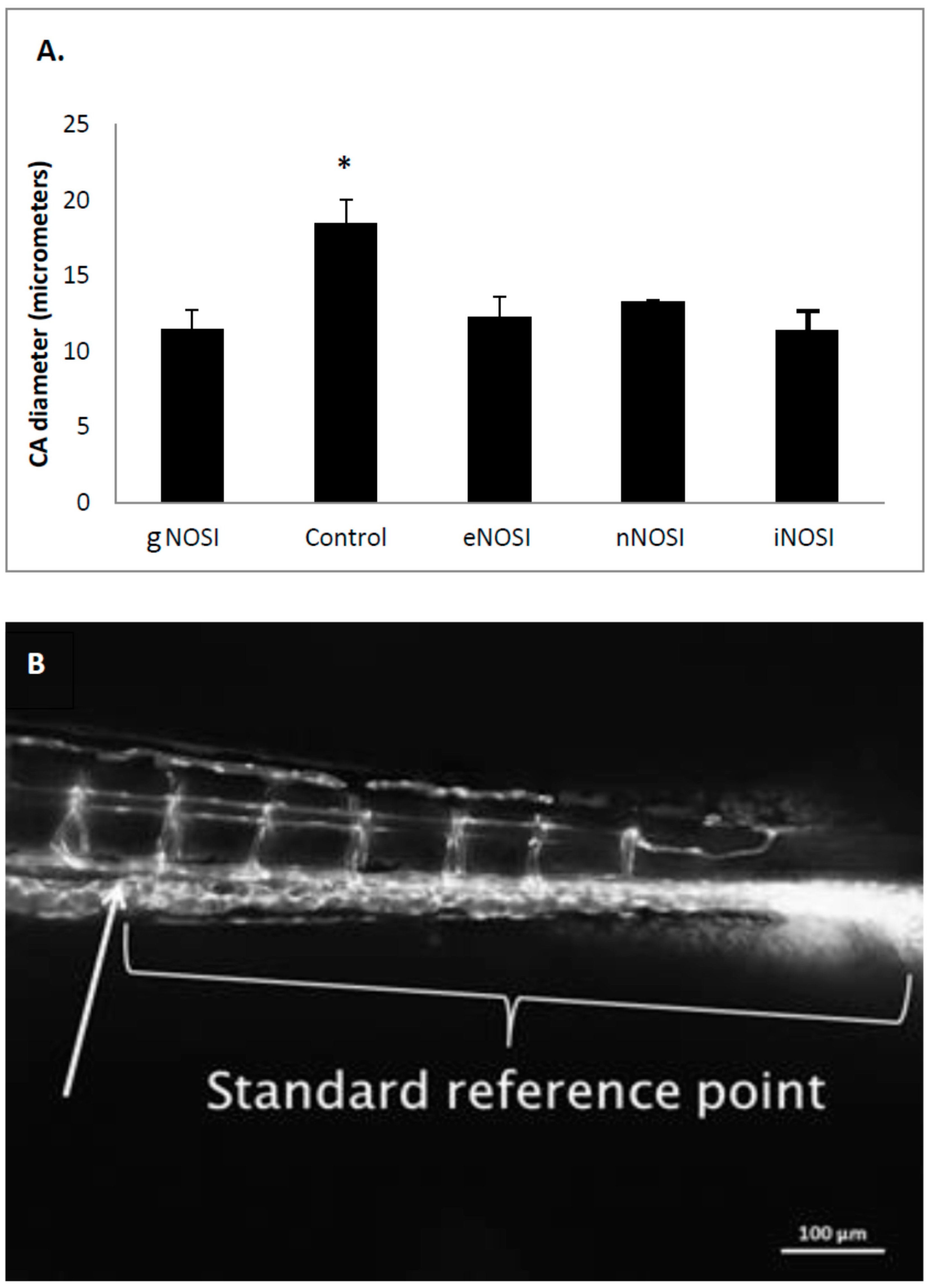
Fig. 7
The effects of various treatments in TG(fli1:EGFP) y1/+y1 (AB) transgenic fish on average tail caudal artery (CA) diameter as measured from confocal z-stack photomicrographs after 4 days of treatment beginning at 48 h post fertilization (hpf). (A) CA diameter is significantly decreased in gNOSI treated fish when compared to that of controls (* p < 0.001). In addition, all three NOSI isoforms also cause a significant decrease in CA diameter (p < 0.05; Bar = ± SD); (B) In order to ensure that measurements of all vessels including the CA were collected in the same location in every confocal- imaged photograph, a standard reference point (arrow) of a distance (875 µm) from the tail region was used. Bar equals 100 µm.