
|
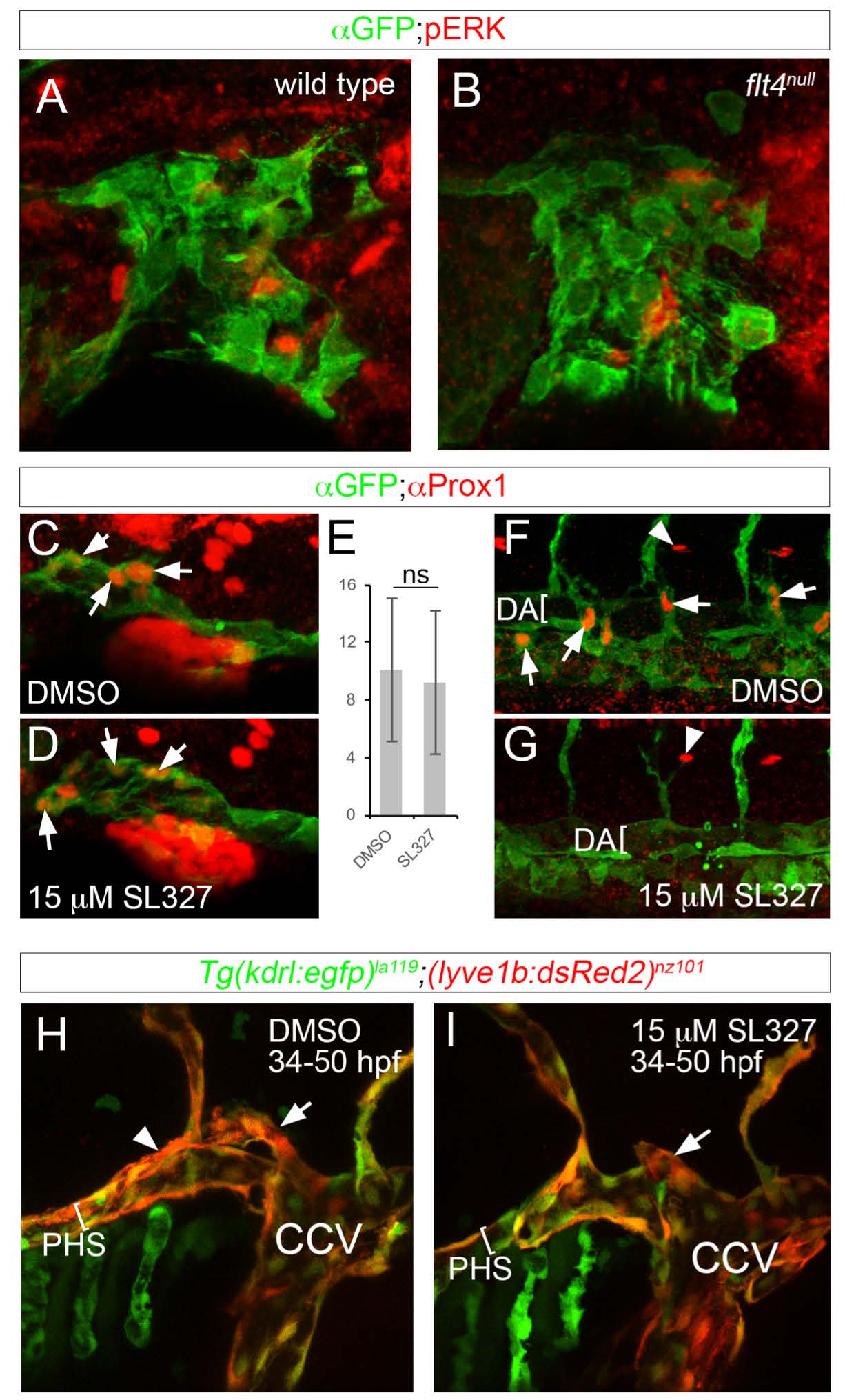
Fig. S4
Flt4 and ERK are dispensable for initial facial lymphatic progenitor specification and sprouting. (A-D, F-I) Confocal images, lateral views, dorsal is up, anterior to the left. (A, B) Tg(fli1a:egfp)y1 of the indicated genotypes immunostained with anti-GFP and anti-pERK antibodies at 36hpf (C, D, F, G) Tg(fli1a:egfp)y1 immunostained with anti-GFP and anti-Prox1 antibodies at 36hpf. (C, F) Embryo treated with DMSO; images from same embryo. (F, G) Embryo treated with 15μM SL327starting at 28hpf; images from same embryo. (E) Average number of Prox1-positive CCV endothelial cells per embryo at 36hpf; ns – not statistically significant, error bars are ± S.D. (H, I) Live Tg(kdr:egfp)la119;(lyve1b:DsRed2)nz101 at 50hpf treated with (H) DMSO or (I) 15μM SL327 starting at 28hpf. Lumen of primary head sinus denoted by a bracket. Position of facial lymphatic vessel derived from PHS is indicated by arrowhead; brach derived from common cardinal vein (CCV) is denoted by an arrow.