
|
Fig. 8
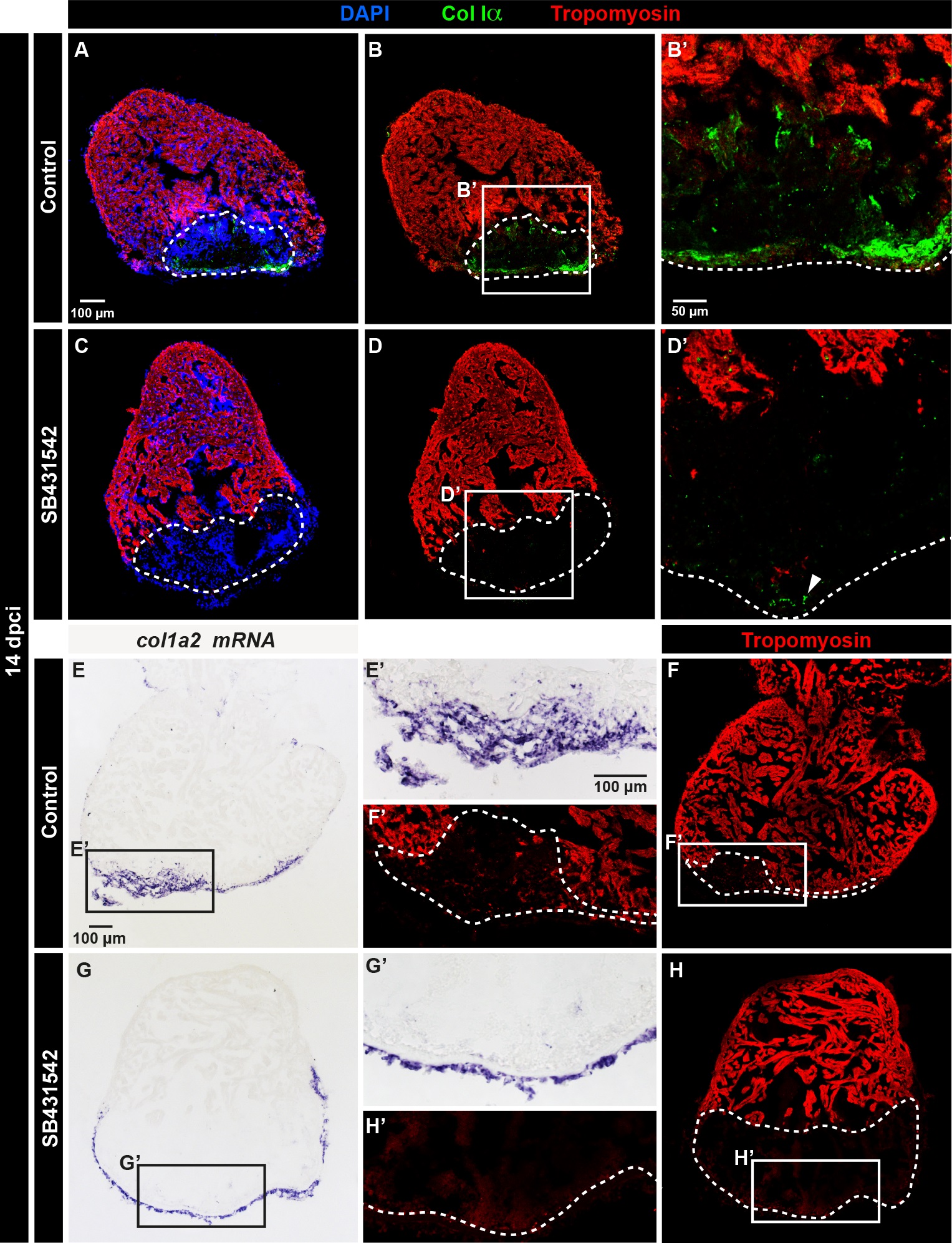
The upregulation of fibrillar Collagen Iα in the post-cryoinjured area is induced by TGF-β signaling.
(A-D) Immunofluorescence staining of heart at 14 dpci using Col Iα (green) and Tropomyosin (red). The post-infarcted tissue is tropomyosin-negative (encircled with a dashed line). (A-B') Control heart displays the presence of fibrillar collagen in the fibrotic tissue. N = 4.(C-D') The treatment with the inhibitor of the TGF-β receptors, SB431542, suppresses Col Iα (green) in the inner wound site. In the epicardium, Col Iα can be still detected (arrowhead). N = 6. (E-H') In-situ hybridization against col1a2 (purple) and immunostaining with Tropomyosin (red). (E-F') Control hearts display expression of col1a2 in the post-infarcted tissue. (G-H') The inhibition of TGF-β signaling with SB431542-treated suppresses col1a2 expression in the inner part of the wound, without affecting the epicardial expression. N = 6.