
|
Fig. S5
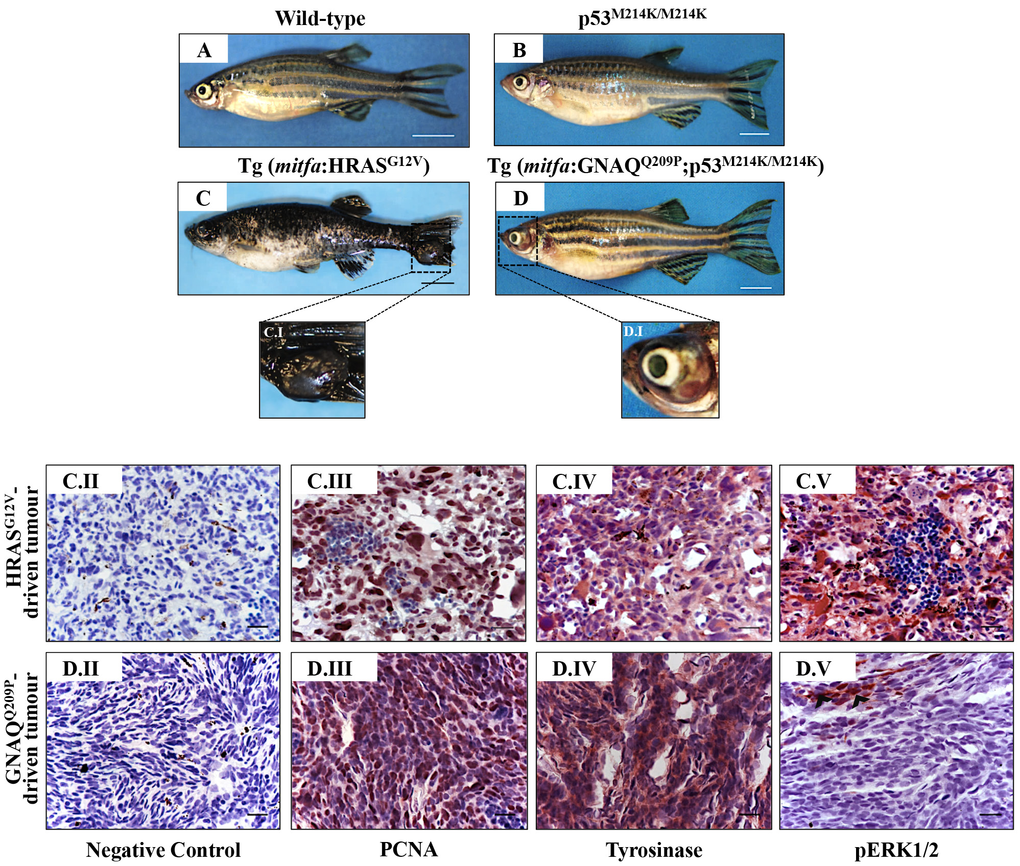
Oncogenic GNAQQ209P and HRASG12V show different potentials in sustaining ERK1/2-MAPK signalling in melanomas of uveal and cutaneous origins. Lateral views of wild-type A. p53M214K/M214K B. Tg (mitfa:HRASG12V) C. and Tg (mitfa:GNAQQ209P;p53M214K/M214K) D. adult zebrafish. Scale bars, 0.5 cm. C.I, D.I. Magnifications of the regions depicted in the black dashed boxes in C and D, respectively. C.II-C.V. Representative IHC images of HRASG12V-driven cutaneous tumours. (C.II) Negative control. (C.III) Numerous melanocytic tumour cells expressing the proliferation marker PCNA: nuclei are stained blue (hematoxylin) and PCNApositive cells acquire dark brown nuclei. (C.IV) Malignant cells expressing the melanocytic differentiation marker tyrosinase. (C.V) Strong immunoreactivity of transformed cutaneous melanocytes to pERK1/2, indicating the activation of ERK1/2-MAPK signalling. D.II-D.V. Representative IHC images of GNAQQ209P-driven uveal tumours. (D.II) Negative control. (D.III) Malignant cells expressing PCNA (dark brown nuclei).(D.IV) Positive immunoreactivity of transformed cells to tyrosinase, indicating the melanocytic origin of the developed ocular neoplasia. (D.V) Transformed uveal melanocytes are barely immunoreactive to pERK1/2, indicating low activity of ERK1/2-MAPK signalling. Abbreviations: PCNA, proliferating cell nuclear antigen. Scale bars, 20 μm.