
Image
|
Figure Caption
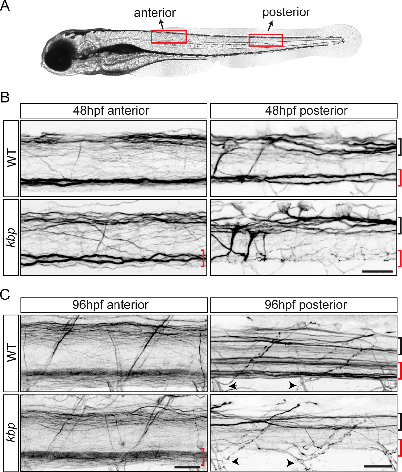
Fig. 1
kbp mutants lack reticulospinal axons in the ventral spinal cord.
A, Location of ‘anterior’ and ‘posterior’ regions analysed. B-C, 3A10 immunostained spinal cords of 48hpf (B) or 96hpf (C) WT and kbp mutant larvae; mutants lack the ventral reticulospinal axons (red bracket), but the dorsal tract (black bracket) and motor axon exit points (arrowhead) appear intact. Scalebars: 25μm.
Figure Data
Acknowledgments
This image is the copyrighted work of the attributed author or publisher, and
ZFIN has permission only to display this image to its users.
Additional permissions should be obtained from the applicable author or publisher of the image.
Full text @ PLoS One