
|
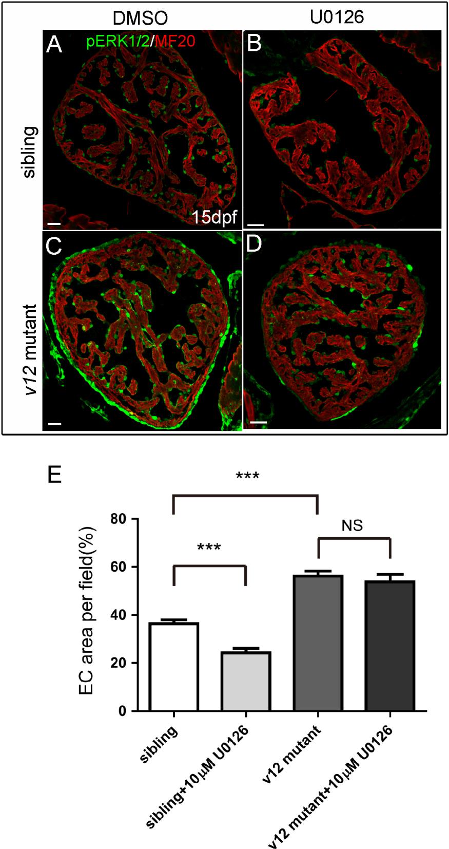
Fig. S10
Coronary vessel density increases in v12 mutant. (A-D) MEK inhibitor (U0126, 10μM) treatment cause partial decrease of the expression level of pERK1/2 in sibling and v12 mutant at 15 dpf. (E) Blood vessel density is presented as percentage of endothelial cell(EC)covered area in a randomly selected area(200µmx 200 µ m).The Tg(fli1a:EGFP) transgenic reporter isused to label endothelial cells. At 40 dpf, blood vessel density is measured in siblings and v12 mutant. v12 mutant has higher blood vessel density than the sibling. MEK inhibitor (U0126, 10μM) treatment impairs coronary development and reduces blood vessel density in wild type, but not in the mutant.