
|
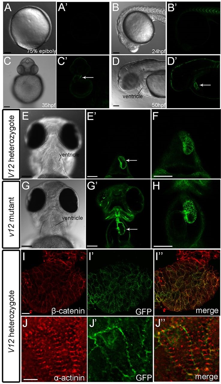
Fig. 1
GFP is highly expressed in the ventricle of v12 zebrafish embryos. (A-D′) GFP is not detected in 75% epiboly embryos (A,A′), and is initially expressed in the somite boundaries at ~24 hpf (B). Cardiac GFP expression starts at ~35 hpf (C,C′) and GFP is highly expressed in the ventricle at 50 hpf (D,D'). Arrows indicate ventricle. (E-H) Ventricular GFP expression in v12 heterozygotes (E-F) and v12 homozygotes (G-H) at 84 hpf shown at high magnification. Arrows indicate ventricle. (I-I'') Immunostaining shows that β-catenin and GFP colocalize close to the cell membrane in v12 heterozygous hearts at 78 hpf. (J-J'') Immunostaining shows that GFP is absent from Z-disc (α-actinin) in v12 heterozygous hearts at 78 hpf. Scale bars: 100 µm in A-D,E-H; 20 µm in I; 5 µm in J.