
|
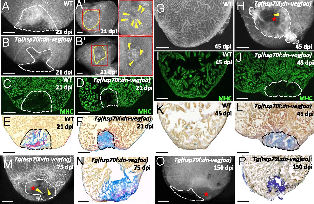
Fig. 5
Blockade of early revascularization prevents vascular invasion and heart regeneration. (A and B) Tg(etv2:EGFP) WT (n = 5) and Tg(hsp70l:dn-vegfaa);Tg(etv2:EGFP) (n = 5) ventricles at 21 dpi. (A¹ and B′) View of the apex of Tg(etv2:EGFP) WT and Tg(hsp70l:dn-vegfaa);Tg(etv2:EGFP) ventricles at 21 dpi. Yellow dotted lines delineate the injured area. Insets (red dotted boxes) show high-magnification images of the injured area. Yellow arrowheads point to coronaries in the injured area. (C and D) Sections of Tg(etv2:EGFP) WT (n = 5) and Tg(hsp70l:dn-vegfaa);Tg(etv2:EGFP) (n = 5) ventricles at 21 dpi. CMs are immunostained with anti-MHC antibody (green). (E and F) Sections of WT (n = 4) and Tg(hsp70l:dn-vegfaa) (n = 5) ventricles at 21 dpi stained with AFOG to identify collagen (blue) and fibrin (red) deposition. (G and H) Tg(etv2:EGFP) WT (n = 5) and Tg(hsp70l:dn-vegfaa);Tg(etv2:EGFP) (n = 4) ventricles at 45 dpi. (I and J) Sections of Tg(etv2:EGFP) WT (n = 5) and Tg(hsp70l:dn-vegfaa);Tg(etv2:EGFP) (n = 4) ventricles at 45 dpi. CMs are immunostained with anti-MHC antibody (green). (K and L) Sections of WT (n = 4) and Tg(hsp70l:dn-vegfaa) (n = 4) ventricles at 45 dpi stained with AFOG to identify scar (blue) and fibrin (red) deposition. (M and O) Tg(hsp70l:dn-vegfaa);Tg(etv2:EGFP) ventricles at 75 (n = 4) and 150 (n = 4) dpi. (N and P) Sections of Tg(hsp70l:dn-vegfaa) ventricles at 75 (n = 4) and 150 (n = 4) dpi stained with AFOG. Dotted lines delineate the injured area. Red stars mark nonvascularized areas. (Scale bars: 100 µm.)