
|
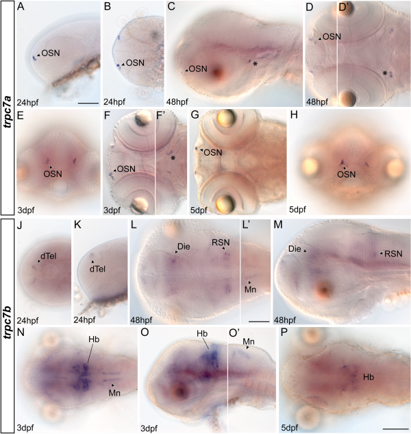
Fig. 8
A-P: Whole-mount in situ hybridization of zebrafish trpc7a (A-H) and trpc7b (J-P). A-D: trpc7a-expressing olfactory sensory neurons (OSNs) in embryos shown laterally (A,C) and dorsally (B,D). Asterisks mark expression domains in the midbrain. The focal plane in D′ differs from D. E,F: Frontal (E) and ventral (F) views on 3 days postfertilization (dpf) whole-mount larvae, F′ shows a different focus to F. Asterisk in F′ labels midbrain cell clusters expressing trpc7a transiently. G,H: Dorsal (G) and frontal (H) views on larvae 5 dpf. J,K: Dorsal (J) and lateral (K) view of the telencephalic cell clusters expressing trpc7b in embryos 24 hr postfertilization (hpf). L,M: Expression of trpc7b as detected in zebrafish 48 hpf shown in dorsal (L) and lateral (M) views. Focus in L′ is more dorsal compared with L. N,O: Expression in 3 dpf larvae, shown in dorsal (N) and lateral (O, O′) views. P: Dorsal whole-mount view on trpc7b expression in larvae 5 dpf. Die, diencephalon; dTel, dorsal telencephalon; Hb, hindbrain; Mn, motoneurons; RSN, reticulospinal neurons. Scale bar = 100 µm in A (applies to all images without scale bar), L′, P.