
Image
|
Figure Caption
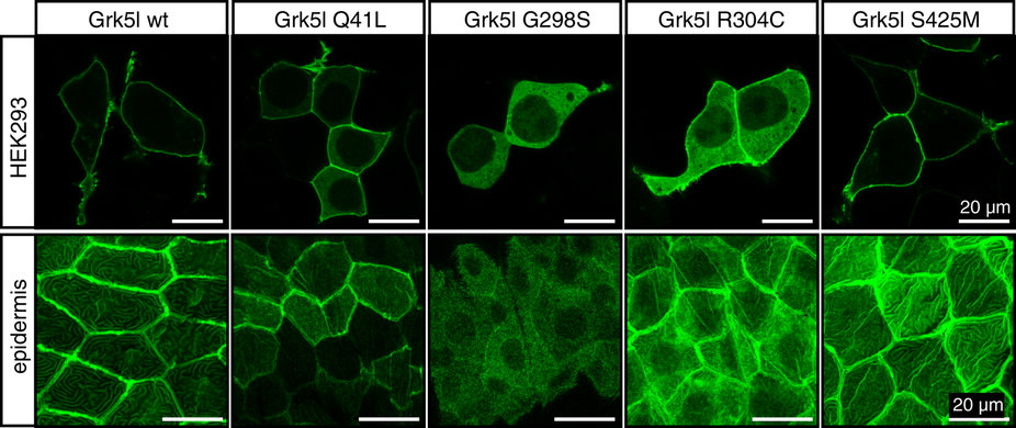
Fig. 3
Subcellular distribution of Grk5l variants.
Upper panel: HEK293 cells were transiently transfected with GFP fusion constructs of wild-type or mutated Grk5l (cloned into pEGFP-N3) and analysed using confocal microscopy. Representative images of three transfections shown. Lower panel: Expression of Grk5l variants in zebrafish produced subcellular distribution of the kinases similar to the results in HEK293 cells. Images show expression in the epidermis. At least five different embryos each from two different injection days were assessed.
Acknowledgments
This image is the copyrighted work of the attributed author or publisher, and
ZFIN has permission only to display this image to its users.
Additional permissions should be obtained from the applicable author or publisher of the image.
Full text @ Sci. Rep.