
|
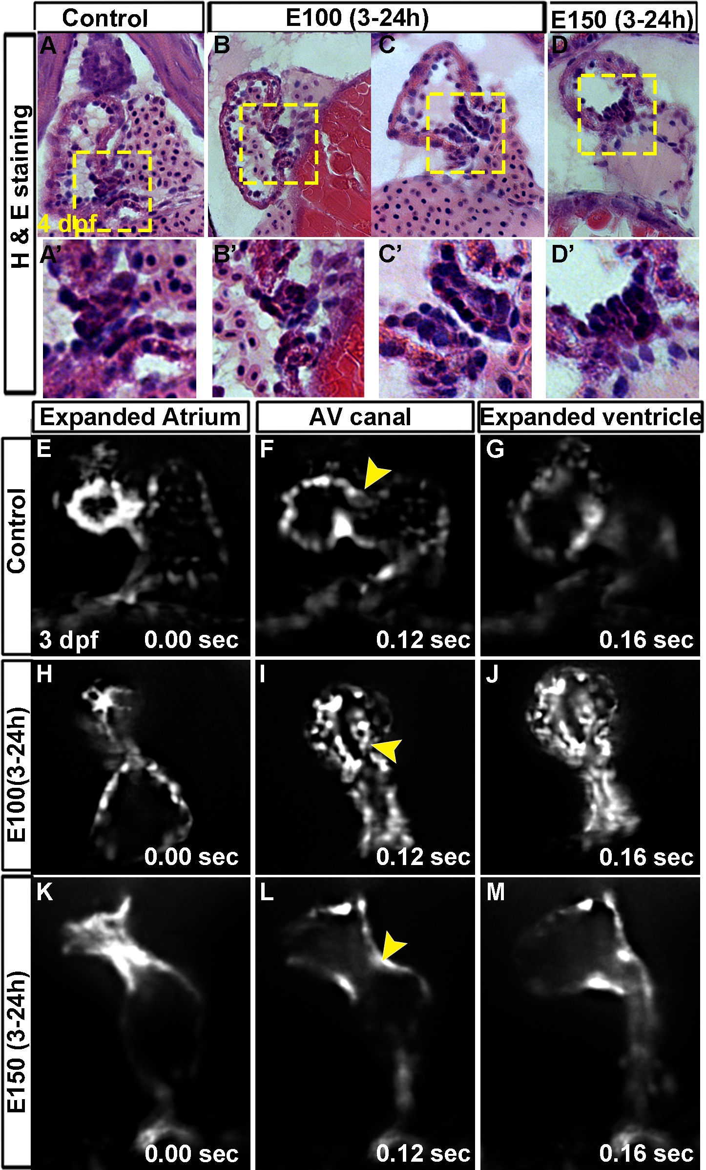
Fig. 1
Embryonic ethanol exposure led to defective AV valve formation in zebrafish.
(A-D′) Hematoxylin and eosin stained coronal sections of 4 dpf zebrafish hearts showed two layers of valve forming cells confined at the AVC in control embryos (A, A′); unorganized clusters of cells at the AV boundary in E100 ethanol treated embryos (B, B′); rows of cells at the extended AVC in E100 ethanol treated embryos (C, C′); and unorganized clusters of cells at the AV boundary in E150 ethanol treated embryos (D, D′). High magnification images of the boxed areas (A′-D′). (E-M) SPIM imaging of beating hearts of Tg(fli1:EGFP) embryos showed completely filled atrium (E, H, K) and ventricular (G, J, M) chambers before ejection of blood in control (E, G) and ethanol treated embryos (H, J, K, M). (F) Control beating heart at the relaxed state showed valve leaflets at the superior and inferior aspects of AVC (F; arrowheads). Ethanol treated embryos showed long valve forming cells extending into the ventricle chamber (E100 ethanol, I) or no valve forming cells at the AVC in E150 treated embryos (L).