
Image
|
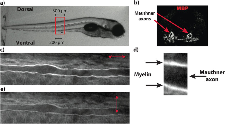
Figure Caption
Fig. 1
P-CARS zebrafish spinal cord model.
(a) The same spinal cord segment can be identified on different imaging sessions using morphological references. (b) The ventral spinal cord contains two Mauthner axons, one on each lateral side (image width: 80 µm). (c) The Mauthner axon can be easily identified with CARS microscopy (image width: 112.5 µm) based on the visualization of (d) its myelin sheaths (image width: 7 µm). (e) Imaging of the same location as in (c) with an orthogonal polarization orientation showing the modulation in CARS intensity. The arrows indicate the orientation of the linear polarization.
Acknowledgments
This image is the copyrighted work of the attributed author or publisher, and
ZFIN has permission only to display this image to its users.
Additional permissions should be obtained from the applicable author or publisher of the image.
Full text @ Sci. Rep.