
|
Fig. 5
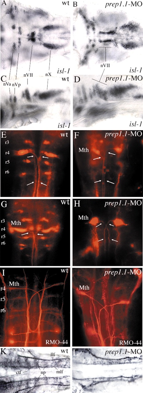
Motor nuclei and ganglia of cranial nerves and reticulospinal neurons, and neuronal architecture of the hindbrain in prep1.1 morphants. (A-D) At 48 hpf, isl1 expression is not affected quantitatively in the motor nuclei of the cranial nerves in prep1.1 morphants (B,D), but the motor neurons of the facial nerve (nVII) have not migrated caudally as in wild-type embryos (A,C). (E-H) Retrograde labelling with rhodamine-dextran of 3-day-old (E,F) and 5-day-old (G,H) embryos. In prep1.1 morphants, only r4 cells are labelled; the projection of their axons (arrows) is similar to that of Mauthner cells but their morphology is slightly different. (I,J) RMO-44 antibody staining of reticulospinal neurons at 48 hpf. Only Mauthner cells are present in prep1.1-MO injected embryos. (K,L) At 30 hpf, the staining pattern of acetylated tubulin antiserum shows that the neuropils and commissural tract fibres, (which are mostly out of focus in wild-type embryos) are much reduced or absent in prep1.1 morphants. Embryos are in dorsal (A,B,E-L) or lateral (C,D) views, with anterior to the left (A-D,K,L) or the top (E-L). ctf, commissural tract fibres; llf, lateral longitudinal fascicle; Mth, Mauthner cells; mlf, medial longitudinal fascicle; np, neuropil; nVa and nVp, anterior and posterior clusters of the trigeminal motor nucleus; nVII and nX, motor nuclei of the facial and vagus nerves, respectively; r, rhombomere; wt, wild-type.