
|
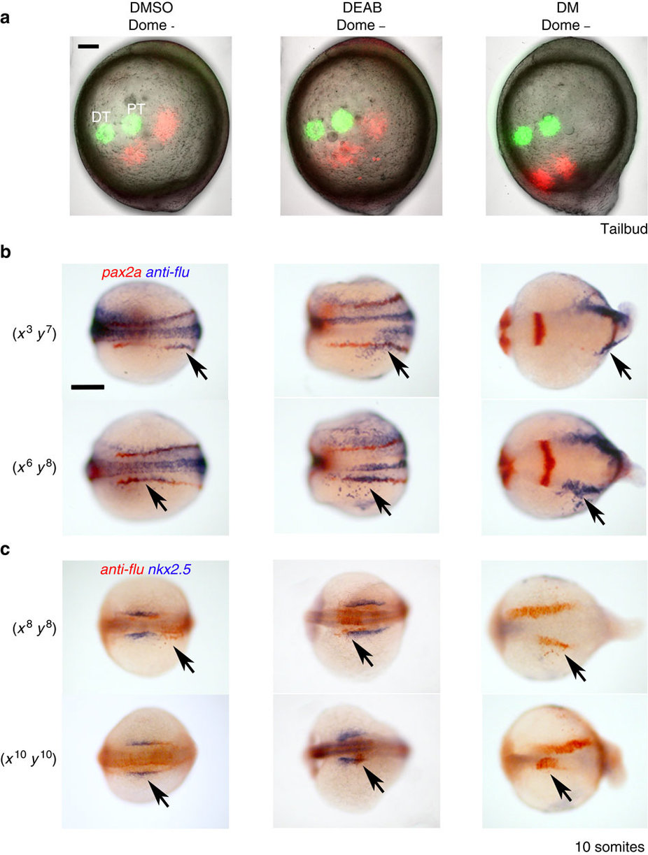
Fig. 8
Effects of BMP and RA on cell fate and cell movement.
(a) Embryos were lineage labelled for distal (DT) and proximal (PT) cells (green) at 85% epiboly. One hour later the positions of these cells were analysed (pseudo-coloured red) and overlayed with the initial lineage labelling. Embryos are lateral views with anterior towards the top. (b) Uncaging cells at (x3, y7) or (x6, y8) at 85% epiboly labelled DT and PT regions, respectively, of the pax2a+ intermediate mesoderm in DMSO- and DEAB-treated 10-somites stage embryos. In DM-treated embryos, (x3, y7) uncaging labelled anterior intermediate mesoderm and a region we predict corresponds to anterior paraxial mesoderm. DM-treated embryos uncaged at (x6, y8) labelled a region of the embryo that likely was composed solely of anterior paraxial mesoderm. Embryos are shown as dorsal views with anterior to the left. (c) Uncaging at (x8, y8) labelled cells just posterior to heart progenitors in DMSO control embryos at the 10-somites stage, but labelled within the posterior domain of expanded heart progenitors (nkx2.5+ cells) in DEAB-treated embryos. Similarly, (x10, y10) labelled heart progenitors in 10-somites stage DMSO control embryos, but mainly labelled the anterior region of the expanded nkx2.5+ domain in DEAB-treated embryos. In DM-treated embryos, heart progenitors were found to sparsely populate the ventral side of the embryo, and (x8, y8) and (x10, y10) labelling at 85% epiboly gave rise to progressively more anterior structures that were positioned close to the dorsal midline. Embryos are shown as dorsal views slightly oblique such that the anterior head region is in view. Scale bars, 100 µm. Arrows indicate uncaged tracer.