
|
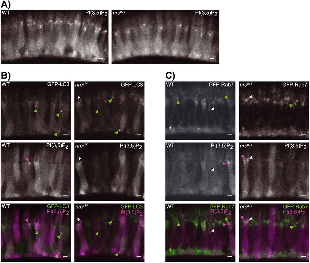
Fig. 5
The distribution of PI(3,5)P2 positive late endosomal compartments is the same in wild type (WT) and nrca14 cells. A; The PI(3,5)P2 probe ML1NX2 localizes to small, punctate structures in the inner segments of WT and nrca14 cone photoreceptors in 5 days post-fertilization Tg(TαCP : mCherry-ML1NX2) transgenic larvae. B: Some PI(3,5)P2 puncta overlap with LC3 positive puncta in nrca14 but not WT cones, agreeing with the decrease in acidic LC3-positive structures observed in Fig. 2. C: The PI(3,5)P2 partially overlaps with Rab7 puncta in both WT and nrca14 cone photoreceptors, but is not found on abnormal Rab7 structures in nrca14 cones. Scale bar = 2 µm in all images.