
|
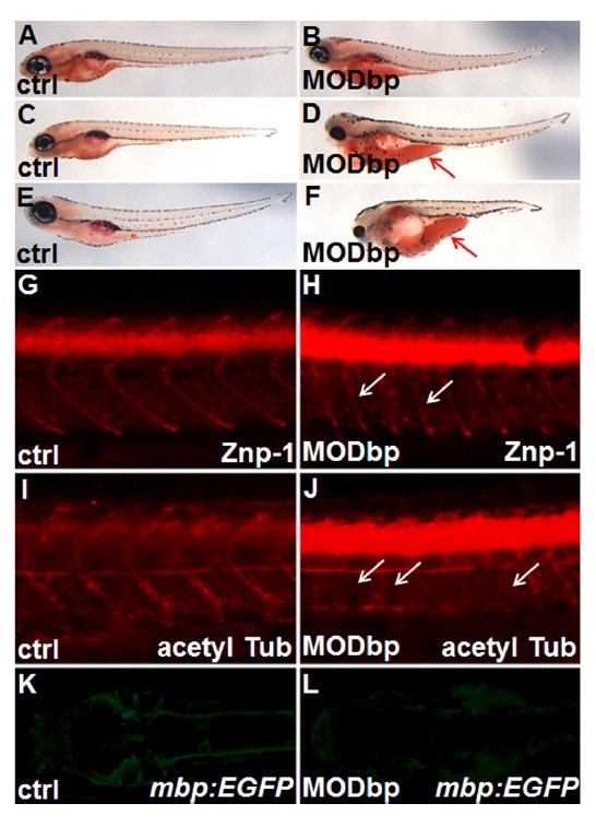
Fig. 3
dbp knockdown affects yolk consumption and neuronal development. (A-F) Embryos with dbp knockdown showed accumulated neutral lipids in the yolk. Oil Red-O staining revealed a significant delay or defect in yolk lipid consumption in dbp knockdown embryos on or after 3 dpf, (A) and (B) (3 dpf); (C) and (D) (4 dpf); (E) and (F) (5 dpf). As compared to control embryo, greater quantities of yolk lipids (visualized in red) were retained in a dbp knockdown embryo. Red arrows in (D) and (F) indicate accumulated lipids in the yolk. (G-L) dbp knockdown impaired neuronal development. Immunostaining using anti-Znp-1 antibody (G, H) or anti-acetylated tubulin (I, J) revealed defective neuronal development, such as discontinued axonal projections [indicated by white arrows in (H)] or suppression of differentiating motor axons [indicated by white arrows in (I)] upon dbp knockdown. (K, L) GFP expression driven by the myelin basic protein (mbp) promoter (indicative of myelinating cells) in an mbp:EGFP transgenic line was significantly reduced following dbp knockdown. Embryos are shown in lateral view with anterior to the left, except in (K) and (L) where embryos are in dorsal view. Partial trunk regions of embryos are shown in (G-J).