
|
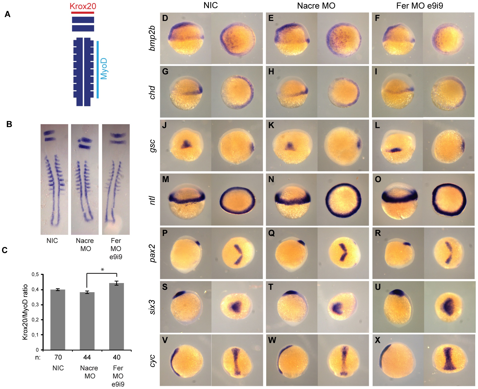
Fig. 4
Fer knockdown resulted in C&E defects but not in changes in cell fate. A. Krox20/myoD in situ hybridization as a method to quantify C&E defects. Krox20 (red) staining for rhombomeres 3 and 5 was used to measure the width, which correlates with convergence cell movements in the embryo. MyoD (light blue) staining for the somites was used to measure the length, i.e. extension of the embryo. The ratio of Krox20/myoD correlates directly with convergence & extension cell movements during gastrulation. B. Flatmounts of krox20/myoD stained NIC (n = 70), control MO (n = 44) and Fer MO (n = 40) embryos. C. The ratio of the width of a krox20-positive rhombomere and the length of 8 somites was determined (* indicates significance, Student′s t-test p<0.005). D-X. Embryos were injected at the 1-cell stage with control MO or Fer e9i9 MO and subjected to ISH for various markers of cell fate determination. Note that the staining of the gsc, pax2, six3 and cyc probes show broader and shorter expression in Fer knockdown embryos than in controls.