
|
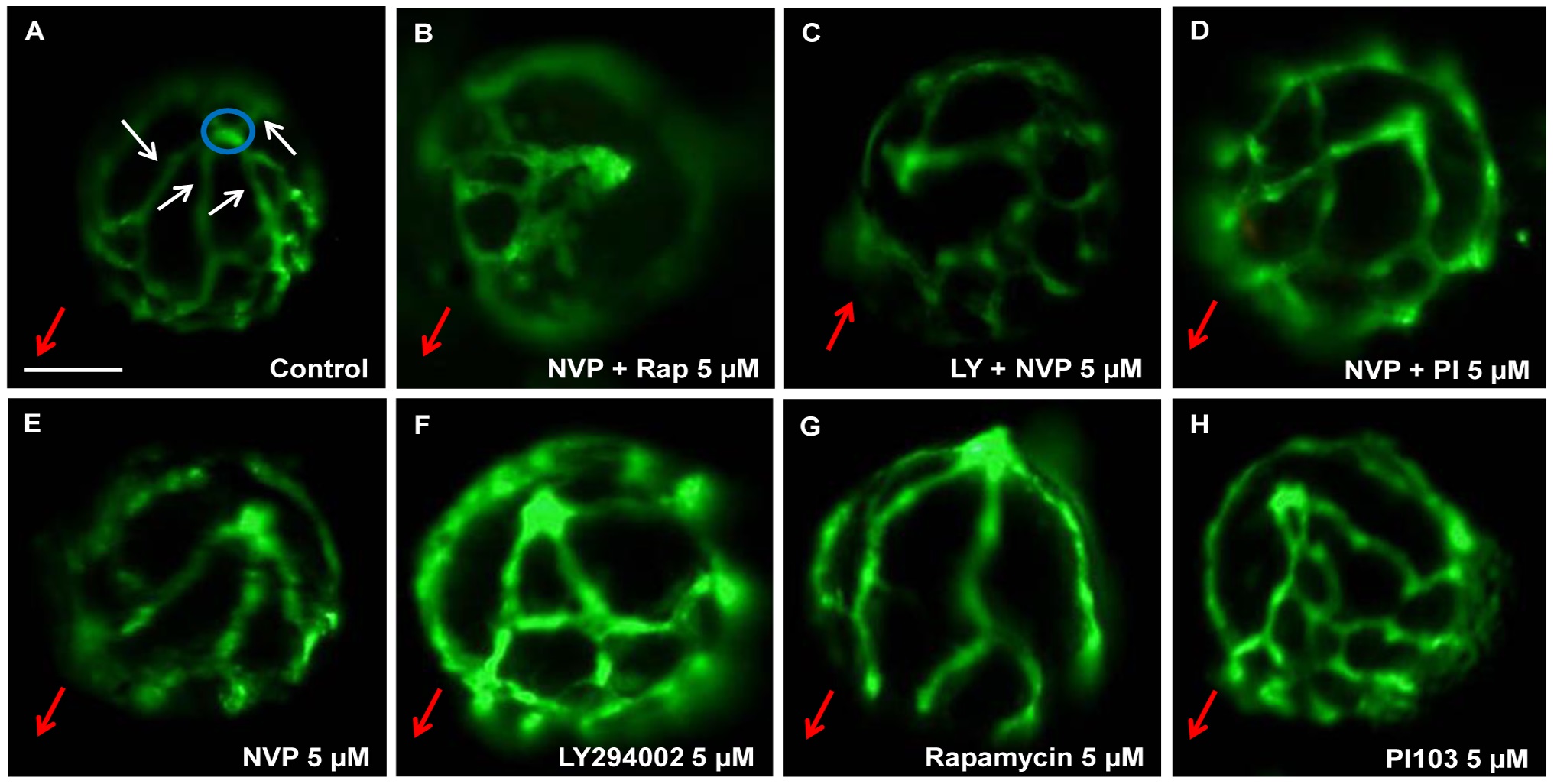
Fig. 5
Qualitative effects of PI3K/Akt/mTOR inhibitors on ocular angiogenesis.
Representative images demonstrating anti-angiogenic effects of PI3K/Akt/mTOR inhibitors on development of HV. 5 µM NVP-BEZ235 + Rapamycin (B), 5 µM LY294002 + NVP-BEZ235 (C) and 5 µM NVP-BEZ235 + PI103 (D) exhibited significant reductions in primary HV branch number compared to 0.1% DMSO-treated larvae (A). Small, but significant differences were observed in the number of primary branches and overall hyaloid vasculature patterning in 5 µM NVP-BEZ235, 5 µM LY294002, 5 µM Rapamycin and 5 µM -PI103-treated larvae (E-H) compared to control (A). Blue circles depict the optic nerve head. White arrows label primary HV branches emanating from the optic disc at the back of the lens. Red arrows indicate the lens orientation pointing in the direction from the optic disk to the lens. Scale bar 100 µm. N = 26-30.