
|
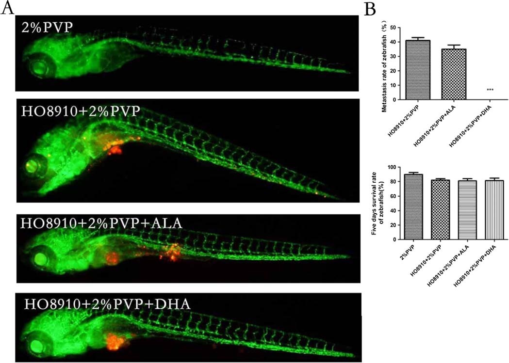
Fig. 5
The effects of ALA and DHA on metastasis of HO8910 cells in model of transgenic zebrafishes. A, 200 cells were injected into the yolk sac of transgenic zebrafishes at 48 hpf per fish, which were pretreated with 120 µM ALA and DHA for 72 hours. A fluorescence microscopy was used to observe dissemination of HO8910 cells in zebrafishes 5 dpi. Green fluorescence shows vasculature of transgenic zebrafish, red fluorescence representing HO8910 cells. B, The metastasis rate and survival rate of zebrafishes was shown by histogram. These experiments were performed in triplicate, and the results showed 1 of 3 independent experiments (***P < 0.005 compared with the control). ALA was used as isotype control. Two percent PVP was used as pharmaceutic adjuvant to prevent cell clumping and needle clogging, and also as a control.