
|
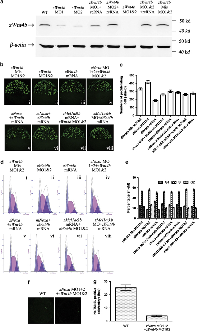
Fig. 6
Mitosis regulated by zNoxa is dependent on zWnt4b. (a) At the shield stage, protein was extracted from embryos and 30 µg of each sample was immunoblotted for zWnt4b and β-actin. β-Actin served as an internal control. (b) Confocal z-stacks of mitotic cells assessed by pH3 staining in embryos which were injected with zWnt4b Mis MO1&2 (i), zWnt4b MO1&2 (ii), zWnt4b mRNA (iii), zNoxa MO1+2 plus zWnt4b MO1&2 (iv), zNoxa mRNA together with zWn4b mRNA (v), mNoxa mRNA plus zWnt4b mRNA (vi), combined zMcl1a&b mRNA and zWnt4b MO1&2 (vii), or zMcl1a&b MO coupled with zWnt4b mRNA (viii) at shield stage. Embryos stained by pH3 were lateral views with animal pole toward the top and dorsal to the right. (c) Quantification of numbers of pH3-positive cells with five embryos for each group in (b). Error bars represent S.E.M. (n=5). (d) A typical FACS analysis of PI-labeled cells in embryos for each group in b. (e) The FACS statistical data of three independent experiments in d. Error bars represent S.E.M. (n=3). (f) Confocal z-stacks of apoptotic cells assessed by TUNEL in embryos that were wt and injected with zNoxa MO1+2 plus zWnt4b MO1&2 at bud stage. Embryos were lateral views with animal pole toward the top and dorsal to the right. (g) Quantification of numbers of TUNEL-positive cells with four embryos for each group in e. Error bars represent S.E.M. (n=4)