
Image
|
Figure Caption
Fig. S4
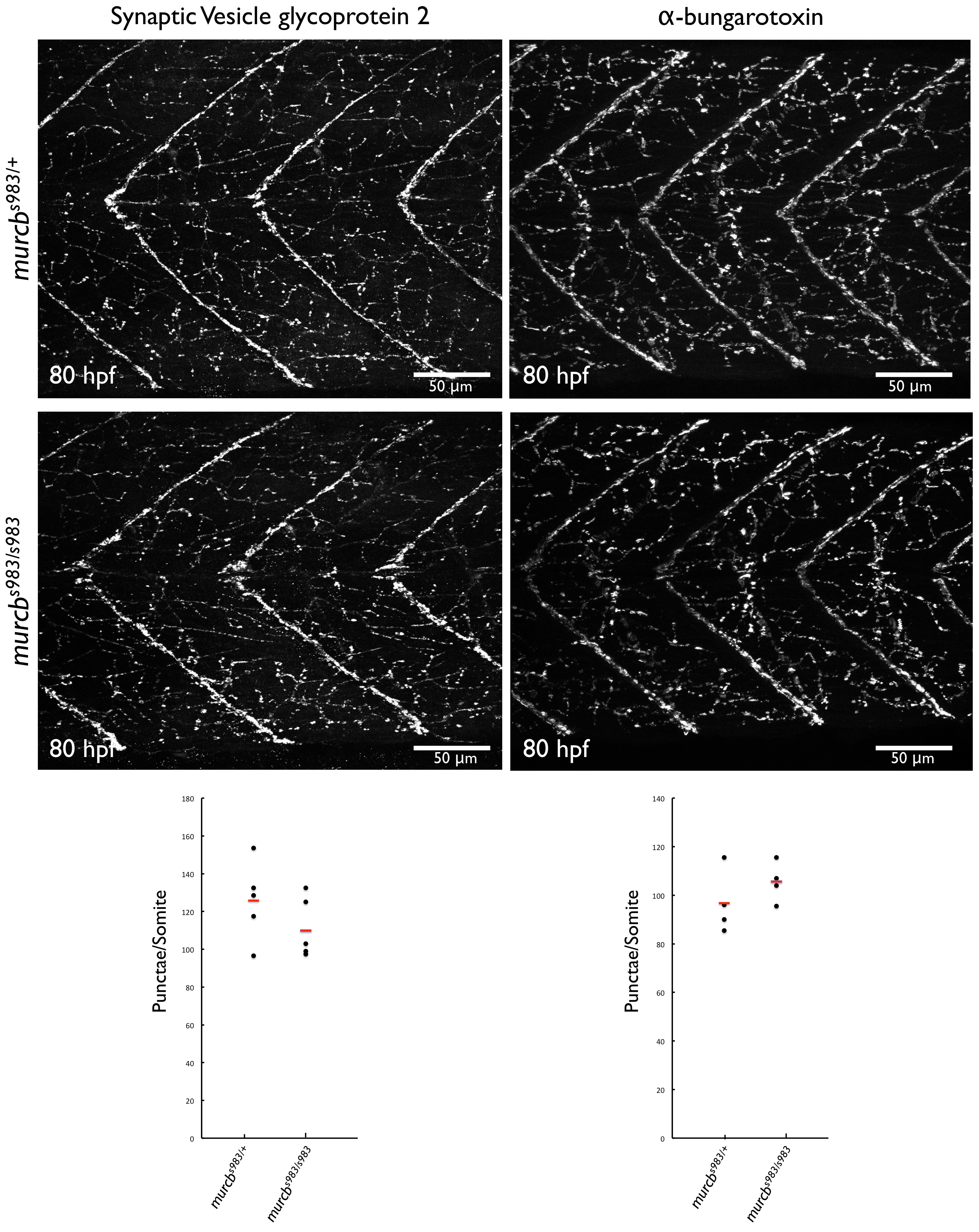
Neuromuscular junction analysis of Cavin4b/Murcb deficient zebrafish.
Representative confocal maximal projections of whole mount Synaptic Vesicle glycoprotein 2 (SV2, left) and α-bungarotoxin (α-BTX, right) staining of the trunk of 80 hpf murcbs983/+ and murcbs983/s983 zebrafish. SV2 immunofluorescence was used to visualize presynaptic structures; α-BTX staining was used to visualize postsynaptic structures. Synaptic punctae per somite were quantified using ImageJ (dotplot, bottom).
Figure Data
Acknowledgments
This image is the copyrighted work of the attributed author or publisher, and
ZFIN has permission only to display this image to its users.
Additional permissions should be obtained from the applicable author or publisher of the image.
Full text @ PLoS Genet.