
|
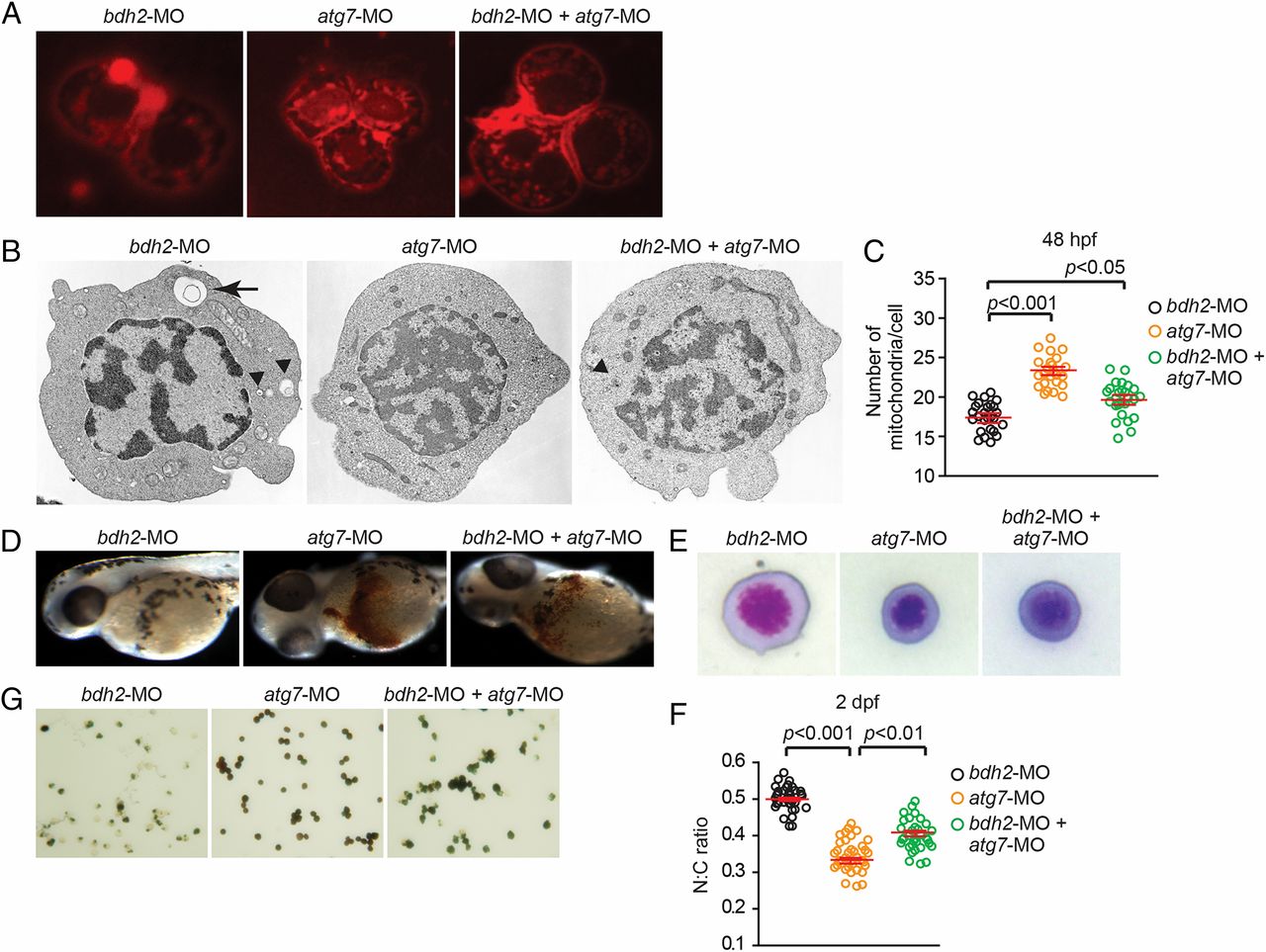
Fig. 5
Suppression of atg7 inhibits mitophagy and rescues hemoglobinization defect in bdh2 morphants. (A) Confocal imaging of MTR-stained circulating erythrocytes from bdh2 or atg7 morphants. Erythrocytes are representative of the mean groups. (B) Representative EM images of erythrocytes isolated from bdh2 or atg7 morphants. (Scale bars: 1 µM.) (C) Total mitochondrial count per cell counted by EM. Mitochondria in autophagosomes were not included in the count. Data are mean ± SD for 50 cells. P < 0.05 was considered significant. (D) Morphological assessment of embryos injected with bdh2 or atg7 MOs. O-dianisidine (O-das) staining confirmed partial restoration of hemoglobinization in atg7-suppressed bdh2 morphants. (E–G) Analysis of circulating erythrocytes from embryos injected with bdh2 or atg7 MOs. (E) May-Grunwald/Giemsa stain (Scale bars: 5 µM.) (F) Tabulation of the N:C area ratio for erythrocytes isolated from embryos injected with bdh2 or atg7 MOs. Coinjection of atg7 MOs decreased the N:C ratio in bdh2 morphants. Data are mean ± SD for 300 cells. P < 0.05 was considered significant. (G) O-dianisidine staining confirmed partial restoration of hemoglobinization in atg7-suppressed bdh2 morphants. Erythrocytes are representative of the mean groups depicted in C. May-Grunwald/Giemsa stain. (Scale bars: 5 µM.)