
Image
|
Figure Caption
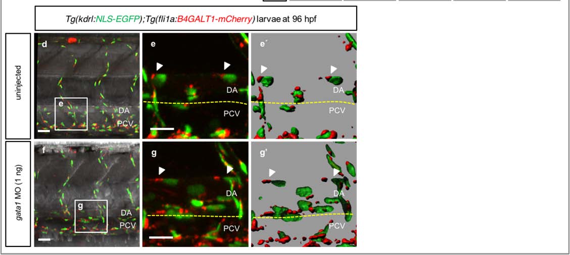
Fig. S4
Endothelial cell polarization is dependent on blood flow.
(a-c) 3D-rendered confocal stack images of the trunk region of a 48 hpf Tg(kdrl:NLS-EGFP);Tg(fli1a:B4GALT1-mCherry) embryo. (b, c) Time-lapse image of ECs identified in (a) (#1, b; #2, c). Surface-rendered images are displayed below. White arrowheads point to polarized ECs, yellow arrowheads to non-polarized ECs. Time (hours:mins) is shown in the top right corner of the images. (d, e, f and g) 3D-rendered confocal stack images of the trunk region of 96 hpf Tg(kdrl:NLS51 EGFP);Tg(fli1a:B4GALT1-mCherry) uninjected (d, e) and gata1 morphant (f, g) larvae. The white boxes in the left panels (d, f) are enlarged in the middle panels (e, g). Surface-rendered images of boxed areas are shown in the right panels (e′, g′). White arrowheads point to polarized ECs. Anterior to the left, dorsal to the top. Scale bars, 20 µm (a, d, e,f and g), 7 µm (b, c). DA, dorsal aorta; PCV, posterior cardinal vein; ISV, intersegmental vessel.
Acknowledgments
This image is the copyrighted work of the attributed author or publisher, and
ZFIN has permission only to display this image to its users.
Additional permissions should be obtained from the applicable author or publisher of the image.
Full text @ Nat. Commun.