
Image
|
Figure Caption
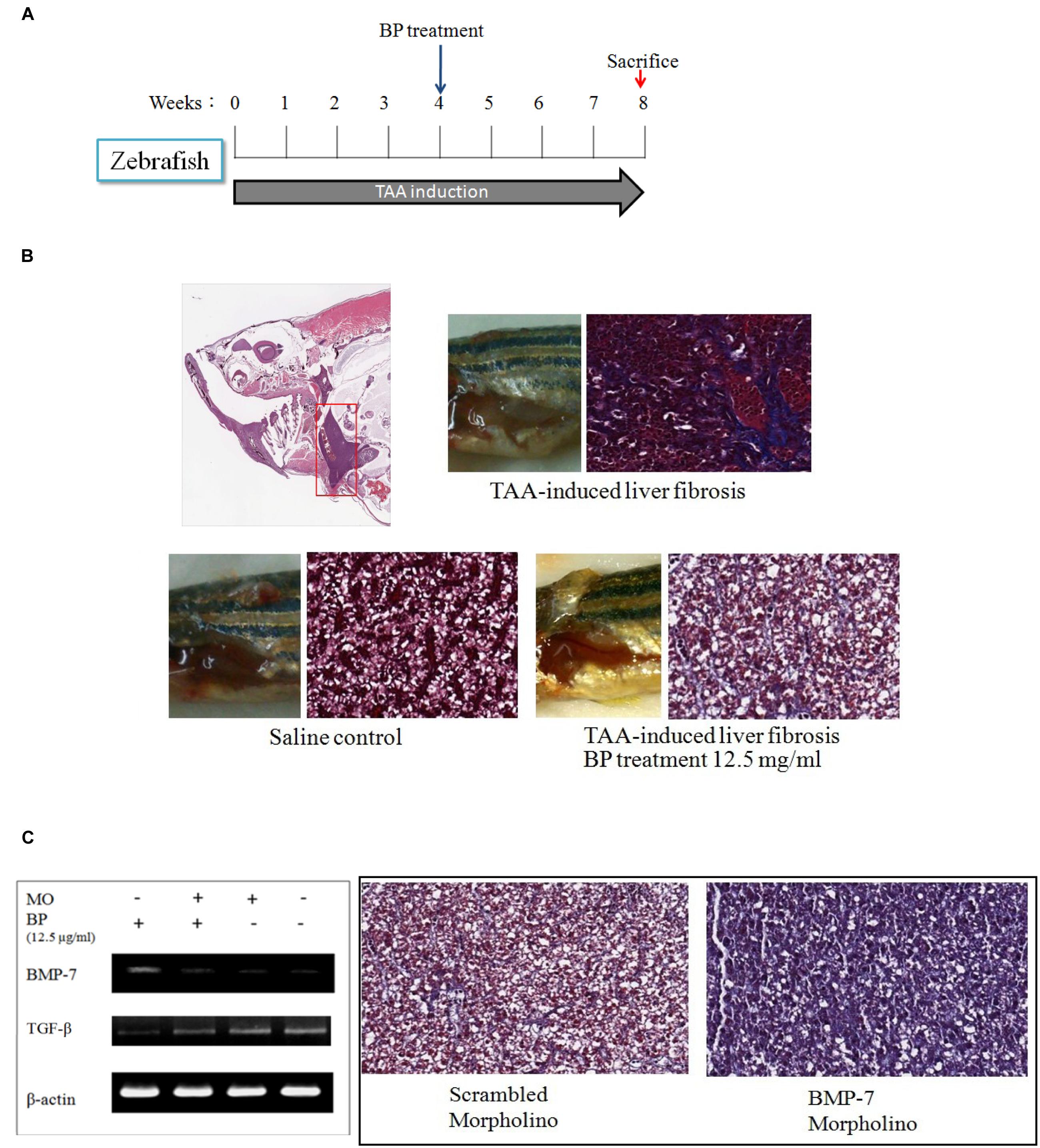
Fig. 5
TAA-induced liver fibrosis in zebra fish. (A) Intraperitoneal injection of TAA (300 mg/kg) three times in a week. Liver tissues were then stained with Masson’s trichrom stain. (B) The collagen content in liver was indicated in blue color. The upper showed TAA-induced liver fibrosis; the lower left showed a normal liver, and lower right showed BP treated TAA-induced liver. (C) TAA-induced zebrafish was injected BMP-7 morpholino (MO) intravenously, and inhibited BP-induced BMP-7 expression (left). Two samples were treated with TAA, BP, and combined with scrambled-morpholino or BMP-7-MO treatment (iv).
Figure Data
Acknowledgments
This image is the copyrighted work of the attributed author or publisher, and
ZFIN has permission only to display this image to its users.
Additional permissions should be obtained from the applicable author or publisher of the image.
Full text @ Front Pharmacol