
|
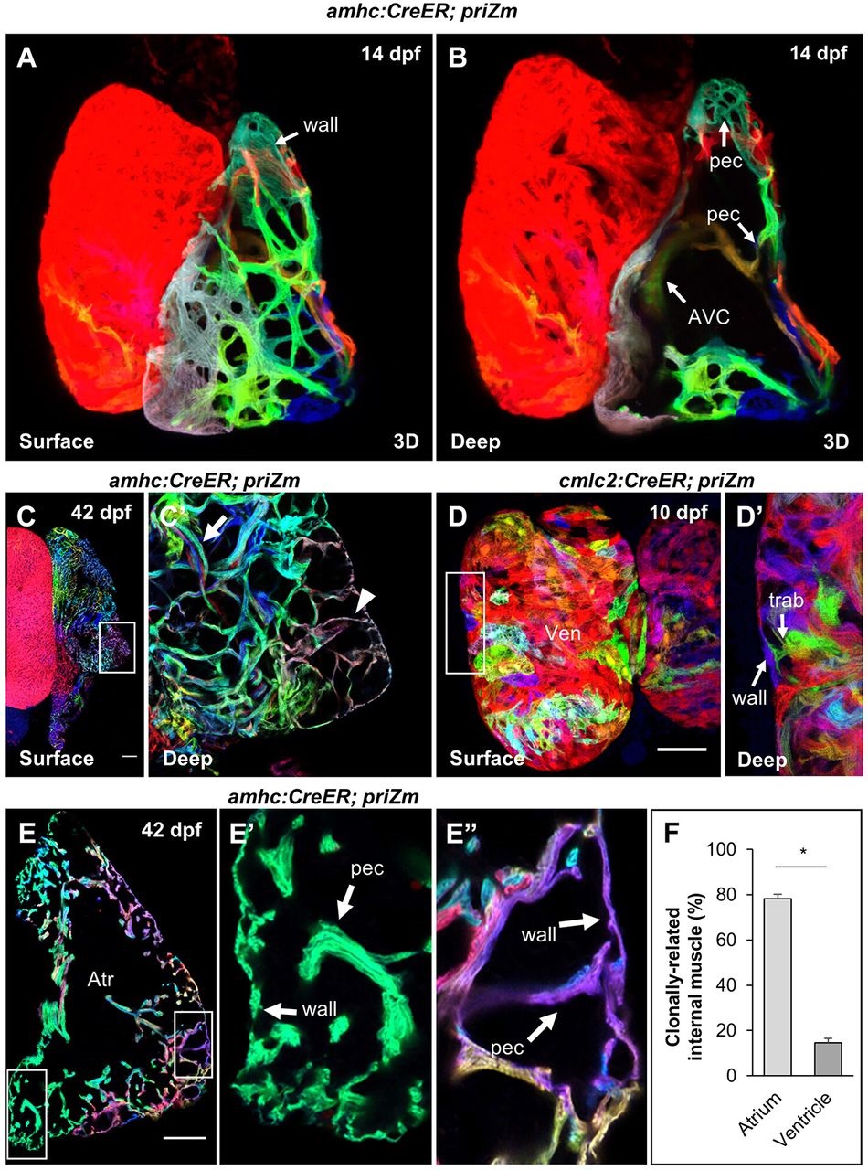
Fig. 2
Atrial pectinate branches directly from wall cardiomyocytes. (A,B) 3D reconstructions of a 14dpf amhc:CreER; priZm heart at the surface (A) and with the anterior hemi-section obscured to show interior structures (B). The atrio-ventricular canal (AVC) and newly formed pectinate fibers (pec) are indicated. (C) Surface and (C′) deep optical sections of a 42dpf atrium. Arrowhead indicates monoclonal pectinate fiber contacting atrial wall. Arrow indicates thicker, polyclonal pectinate fiber deeper in the lumen. (D) Surface and (D2) deep optical sections of a 10dpf cmlc2:CreER; priZm ventricle treated with 4-HT at 3dpf. Arrows indicate a point of contact between a trabecular (trab) and ventricular wall cardiomyocyte (wall). Note that red cardiomyocytes, as the unrecombined color, are uninformative for clonal concordance assessment. (E) Cryosection of a 42dpf amhc:CreER; priZm atrium exhibiting monoclonal, clonally concordant pectinate (pec) muscle (E′) and a polyclonal fiber containing clonally concordant muscle (E′′); Atr, atrium. (F) Frequency with which pectinate and trabecular muscle share a clonal origin with adjacent cardiomyocytes of the atrial and ventricular wall, respectively [means±s.e.m.; n=5 (atria) and 11 (ventricles) from 16 hearts]. Asterisk indicates significantly different means (two-tailed Student′s t-test, P<0.001). Scale bars: 100µm in C,C′,E-E′′ and 50µm in D,D2.