
|
Fig. 4
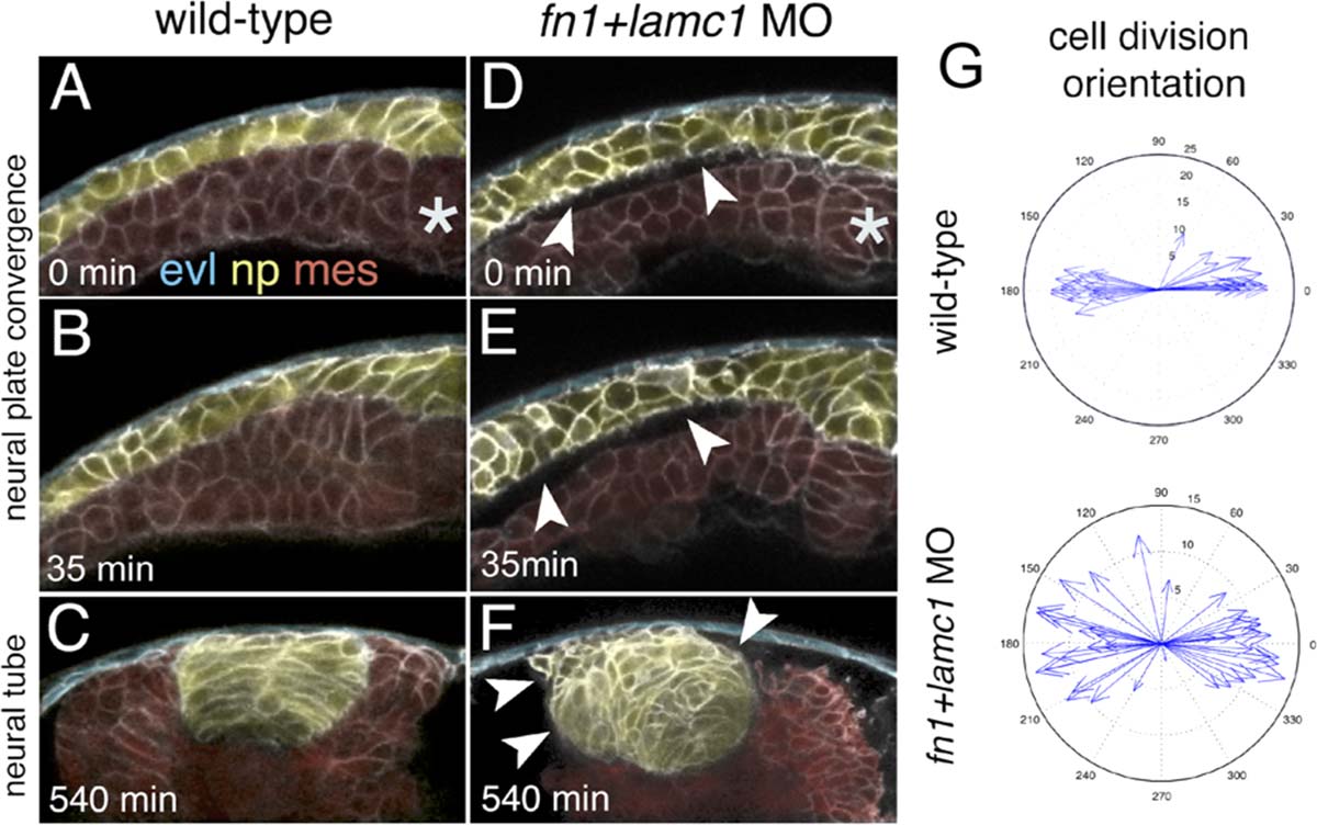
ECM is required to maintain close tissue apposition of neural tissue and mesoderm. A-C: Selected frames from a time-lapse sequence showing close apposition of mesoderm and neural tissue throughout neurulation in wild-type embryo. There is no space apparent between mesoderm and neural tissue. By 20 hpf (540 min) the neural tube and mesoderm remains closely apposed. D-F: Time-lapse sequence from an Fn1/LamC1 deficient embryo showing the presence of gaps between the neural plate and mesoderm from early time point of neurulation (arrowheads). By 20 hpf, tissue gaps are still present (arrowheads) and neural tube architecture becomes impaired and no clear midline is seen. In A-F, the enveloping layer (evl) has been pseudocolored in blue, neural plate (np) has been pseudocolored in yellow, and the mesoderm (mes) has been pseudocolored in red. G: Analysis of cell division orientation and location shows the normal midline location of divisions is lost and stereotyped mediolateral orientation of divisions is disrupted in Fn1/LamC1 deficient embryos. hpf, hours post fertilization; min, minutes and asterisks mean embryo dorsal midline.