
|
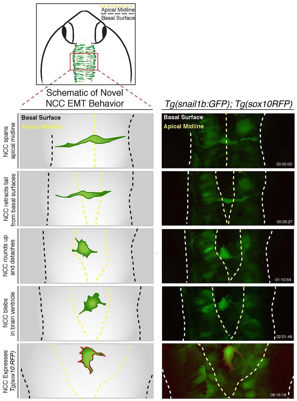
Fig. 3
Dorsal midline neural epithelial progenitor cells display novel delamination behaviors of EMT. Schematic illustrating imaging area (top) and novel delamination behavior of a dorsal midline neuroepithelial progenitor (left); the schematic is derived from confocal time-lapse images of Tg(snai1b:GFP); Tg(sox10:RFP) embryos (right). Images show a GFP-positive cell initially spanning the dorsal midline before contracting apical attachments from each side of the neuroepithelium. Subsequently, GFP-positive cells detach and become rounded with filopodia and blebbing protrusions, and ultimately express sox10-mRFP at cell membranes. See also Movie 3.