
Image
|
Figure Caption
Fig. S2
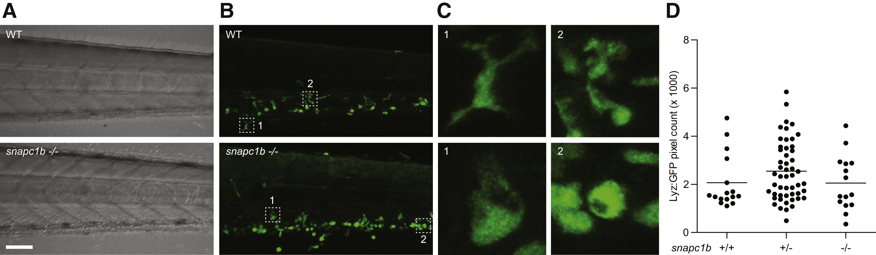
snapc1b Mutants Have Numerous Vacuolated Macrophages and Normal Neutrophil Numbers in the Caudal Hematopoietic Tissue, Related to Figure 1
(A and B) (A) Brightfield and (B) confocal images of the CHT of representative WT and snapc1b-/- mutant larvae at 5 dpf. Scale bar 50µm.
(C) 8X magnification of outlined regions in (B) showing normal (top) and vacuolated (bottom) morphology.
(D) Quantification of Lyz:eGFP positive, green fluorescent neutrophils in snapc1b+/- incross larvae at 6 dpf.
Acknowledgments
This image is the copyrighted work of the attributed author or publisher, and
ZFIN has permission only to display this image to its users.
Additional permissions should be obtained from the applicable author or publisher of the image.
Reprinted from Cell, 165, Berg, R.D., Levitte, S., O'Sullivan, M.P., O'Leary, S.M., Cambier, C.J., Cameron, J., Takaki, K.K., Moens, C.B., Tobin, D.M., Keane, J., Ramakrishnan, L., Lysosomal Disorders Drive Susceptibility to Tuberculosis by Compromising Macrophage Migration, 139-152, Copyright (2016) with permission from Elsevier. Full text @ Cell