
|
Fig. 6
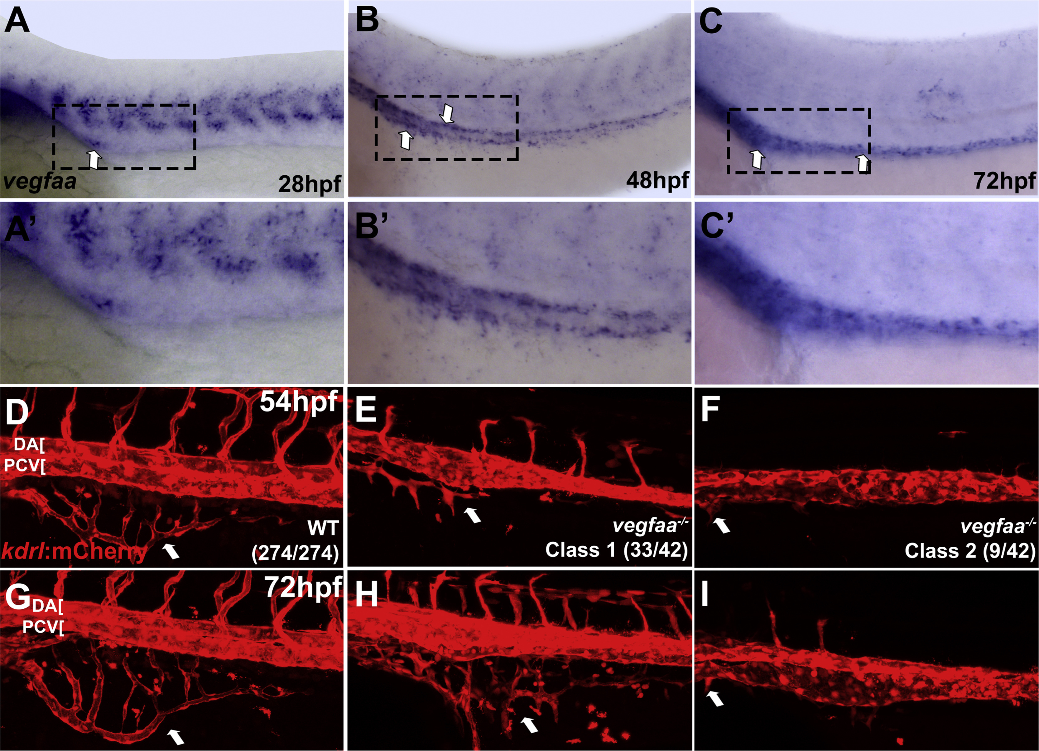
vegfaa provides a candidate guidance signal required for intestinal vessel development. In situ hybridization of wild type embryos (A-C′) and confocal images of live Tg(kdrl:mCherry) embryos (D-F′). (A-C) vegfaa is expressed at 28, 48, and 72 hpf in a narrow stripe of cells within the endoderm along the yolk and yolk extension in the same region where angioblasts coalesce into the SIA and SIV. (A′-C′) show higher magnification of the embryos in A-C. (D-F) vegfaa-/- embryos at 54 hpf exhibit strong inhibition (classs 1) or complete absence of SIV development (class 2). In addition, single axial vessel and inhibition of intersegmental vessel angiogenesis, where ISVs are absent or do not extend past the midline, are observed. (D′-F′) The same embryos at 72 hpf show little to no recovery of more severe defects. (Class 1 - moderate SIV defect, Class 2 - absent or nearly absent SIV). Arrows indicate SIV. Anterior-left, dorsal-top.
Reprinted from Developmental Biology, 411(1), Koenig, A.L., Baltrunaite, K., Bower, N.I., Rossi, A., Stainier, D.Y., Hogan, B.M., Sumanas, S., Vegfa signaling promotes zebrafish intestinal vasculature development through endothelial cell migration from the posterior cardinal vein, 115-27, Copyright (2016) with permission from Elsevier. Full text @ Dev. Biol.