
Image
|
Figure Caption
Fig. S5
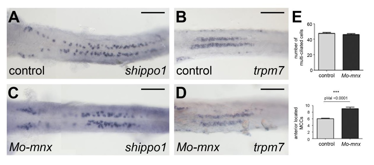
Ciliated cell specification occurs independently of Mnx functions. Expression of multiciliated and transporting epithelial cell markers is shown for 2dpf embryos (ventral view). Multi-ciliated marker shippo1/odf3b (A, C) and mono-ciliated marker trpm7 (B, D) are comparable in controls and mnx morpholino injected embryos. Scale bars correspond to 100µm. Cell numbers are summarized in (E).
Acknowledgments
This image is the copyrighted work of the attributed author or publisher, and
ZFIN has permission only to display this image to its users.
Additional permissions should be obtained from the applicable author or publisher of the image.
Reprinted from Developmental Biology, 411(1), Ott, E., Wendik, B., Srivastava, M., Pacho, F., Töchterle, S., Salvenmoser, W., Meyer, D., Pronephric tubule morphogenesis in zebrafish depends on Mnx mediated repression of irx1b within the intermediate mesoderm, 101-14, Copyright (2016) with permission from Elsevier. Full text @ Dev. Biol.