
Image
|
Figure Caption
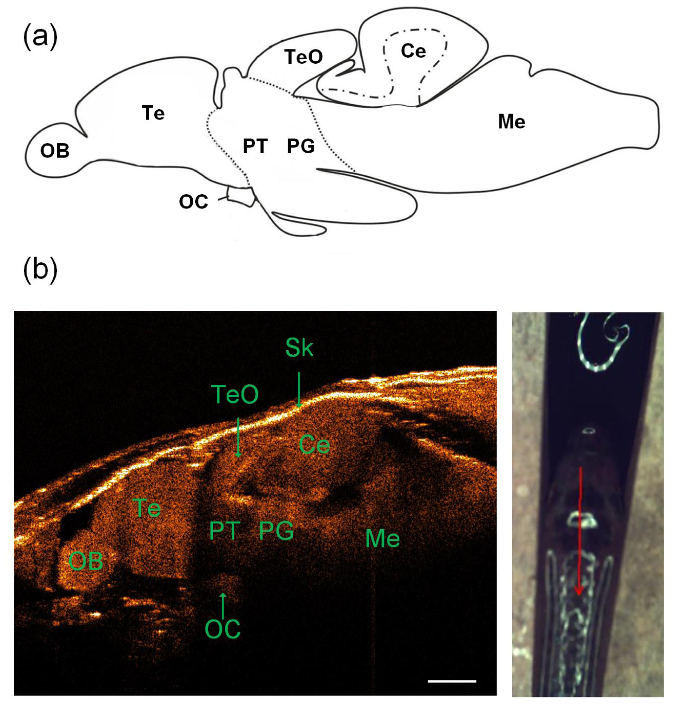
Fig. 2
(a) Schematic of the lateral view of adult zebrafish brain (the top row) [27]. (b) A representative sagittal SD-OCT image of the adult zebrafish brain (bottom left) along the red profile as shown in the photograph (bottom right): OB, olfactory bulb; OC, optic commissure; Te, telencephalon; TeO, tectum opticum; Ce, cerebellum; Me, medulla; PG, preglomerular complex; PT, posterior tuberculum; Sk, skull. The scale bar is 500 µm.
Acknowledgments
This image is the copyrighted work of the attributed author or publisher, and
ZFIN has permission only to display this image to its users.
Additional permissions should be obtained from the applicable author or publisher of the image.
Full text @ Biomed. Opt. Express