
|
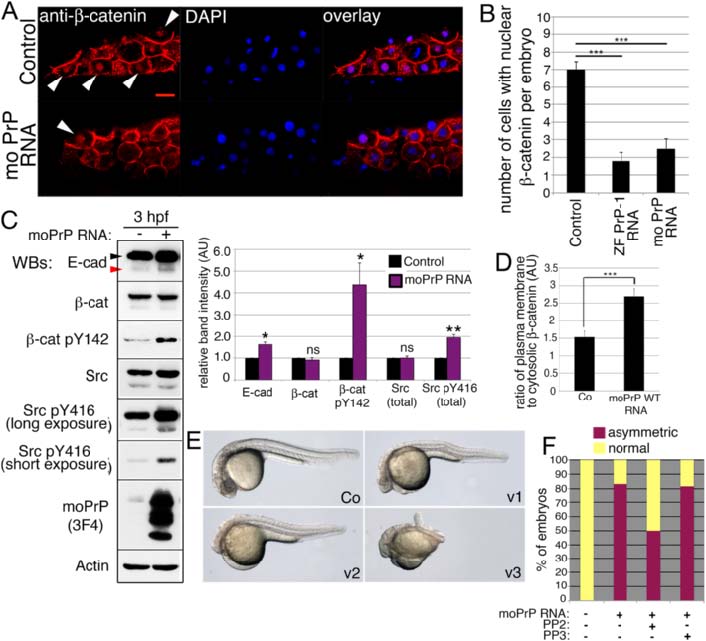
Fig. 6
PrP overexpression up-regulates E-cadherin, inhibits nuclear β-catenin and produces ventralized embryos. a. Localization of β-catenin in the nuclei of dorsal marginal blastomeres at 3 hpf (immunofluorescence). Arrowheads indicate β-catenin-positive nuclei. Scale bar = 20 µm. b. Quantification of cells with nuclear β-catenin in 3 hpf embryos expressing zebrafish and mouse WT PrP constructs. Average numbers of cells with nuclear β-catenin per embryo ± SEM are shown (n = 10, three independent experiments); statistical significance was assessed using unpaired, two-tailed t-tests; *** = p ≤ 0.001. c. Levels of E-cadherin, β-catenin and SFKs in 3 hpf embryos expressing mouse PrP (Western blot and densitometric analysis). E-cadherin arrowheads as in Fig. 1. Densitometric analysis of Western blot bands expressed in arbitrary units (AU); average values of three independent experiments ± SEM are shown; statistical significance was assessed using unpaired, two-tailed t-tests; ns = not significant (p > 0.05), * = p ≤ 0.05, ** = p ≤ 0.01. d. Ratios of plasma membrane to cytosolic β-catenin immunofluorescence in dorsal blastomeres of 3 hpf embryos. Mean ratios ± SEM are from 10 cells/embryo (n = 7, three independent experiments); statistical significance assessed by unpaired, two-tailed t-test, *** = p < 0.001. e. Ventralized hypomorphic phenotypes (v1-3) of 1 dpf embryos injected with 20 ng/µl mouse PrP mRNA. f. Quantification of 6 hpf embryos with abnormal dorsoventral specification upon injection of mouse PrP alone, together with PP2, or with its inactive analog PP3