
|
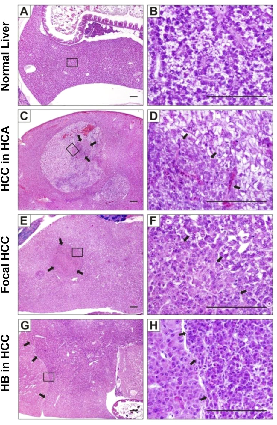
Fig. 4
Heterogeneous liver tumors induced by krasV12.
Histopathological examinations of liver tumors from Triple-Tg fish induced at 1-month-old. (A,B) Representative normal liver section from Triple-Tg fish without induction. (C,D) Liver tumor from induced Triple-Tg fish after 15 weeks displayed many vacuolated hepatocellular adenoma (HCA) with carcinoma grade 2 (HCC) arising in the center. (E,F) Liver tumor from induced Triple-Tg fish after 19 weeks showed HCC (grade 1) occupying 80% of the liver volume, with HCC (grade 2) arising centrally. (G,H) A Triple-Tg fish at 28 weeks after induction showing HCC (grade 2–3) involving the entire liver with extensive areas of hepatoblastoma (HB). Right panel showed high magnification of boxed area in the left panel. Arrows indicated the boundaries between different types of liver tumors. Scale bars, 100 µm.