
Image
|
Figure Caption
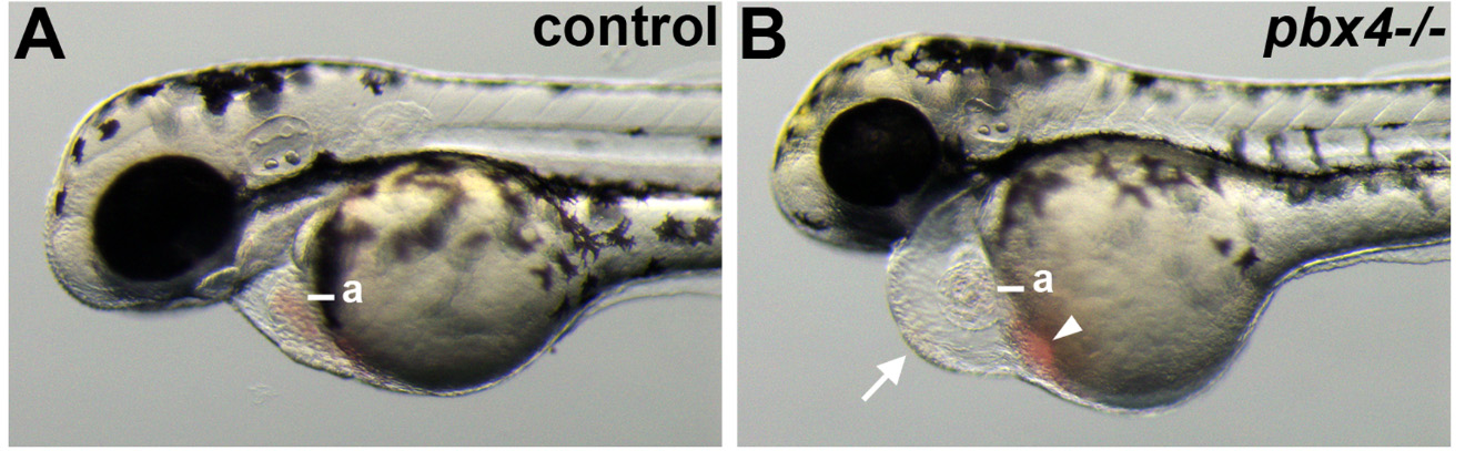
Fig. 1
Defective heart development in pbx4 mutant embryos. (A,B) Lateral views of live (A) control and (B) pbx4b557-/- embryos at 50 h post fertilization (hpf). pbx4b557 mutant embryos show pericardial edema (arrow in B) and blood pooled near the atrium (arrowhead in B). a, atrium. For controls, n = 23. pbx4b557 mutant embryos (n = 20) all show similar phenotypes as in (B). Anterior is to the left.
Figure Data
Acknowledgments
This image is the copyrighted work of the attributed author or publisher, and
ZFIN has permission only to display this image to its users.
Additional permissions should be obtained from the applicable author or publisher of the image.
Full text @ J Dev Biol