
|
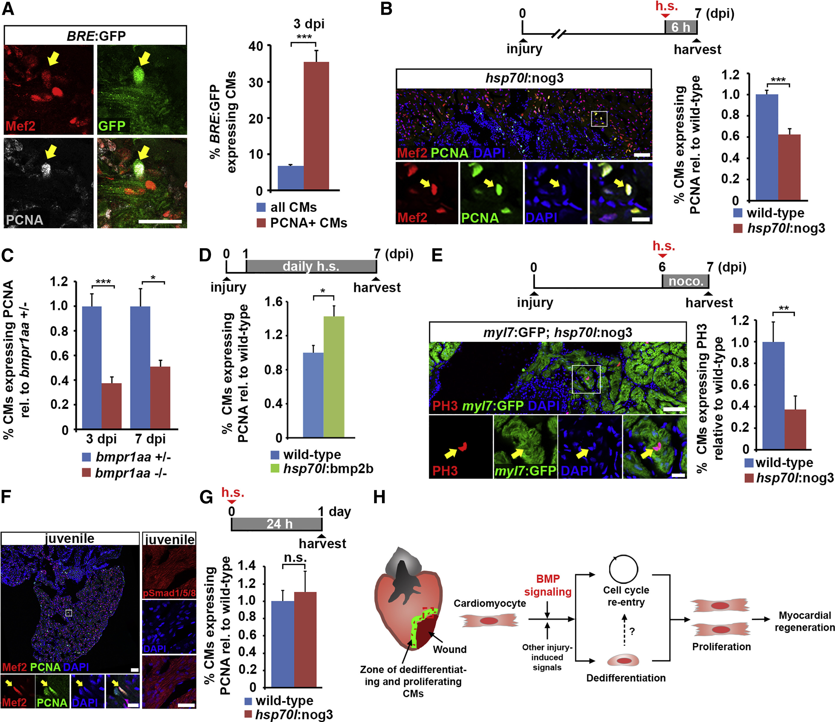
Fig. 7
BMP Signaling is Required for Injury-Induced but Not Physiological Cardiomyocyte Proliferation
(A) Co-localization of Mef2 and PCNA with GFP (arrow), which reports BMP-mediated transcriptional activity in BRE:GFP transgenic hearts, at 3 dpi. Average percentages of PCNA+ and of all cardiomyocytes that express GFP are plotted. Error bars represent SEM. Student’s t test, ***p = 0.000023.
(B) Expression of noggin3 in hsp70l:nog3 transgenics for 6 hr is sufficient to reduce PCNA expression in cardiomyocytes (identified by nuclear Mef2, arrow) at the wound border at 7 dpi. Boxed area is shown as magnified view. Average percentage of cardiomyocytes expressing PCNA relative to wild-type is plotted. Error bars represent SEM. Student’s t test, ***p = 0.00095. Scale bar, 50 µm.
(C) Hearts homozygous for a loss-of-function mutation of the BMP type-I receptor bmpr1aa display reduced expression of PCNA in cardiomyocytes (identified by nuclear Mef2) at the wound border at 3 and 7 dpi compared with bmpr1aa+/- hearts. Average percentage of cardiomyocytes expressing PCNA relative to bmpr1aa+/- control is plotted. Error bars represent SEM. Student’s t test, ***p = 0.0002 (3 dpi), *p = 0.01 (7 dpi).
(D) Overexpression of bmp2b for 6 days via daily heat shock is sufficient to increase PCNA expression in cardiomyocytes at the wound border in hsp70l:bmp2b transgenics relative to heat-shocked wild-type siblings at 7 dpi. Average percentage of cardiomyocytes expressing PCNA relative to wild-type is plotted. Error bars represent SEM. Student’s t test, *p = 0.011.
(E) Expression of noggin3 for 24 hr in hsp70l:nog3; myl7:GFP double transgenics that were treated with 5 µM nocodazole reduces the percentage of phospho-Histone 3 (PH3)+ mitotic cardiomyocytes (arrow). Boxed area is shown as magnified view. Average percentage of cardiomyocytes expressing PH3 relative to wild-type is plotted. Error bars represent SEM. Student’s t test, **p = 0.0068. Scale bar, 50 µm (overview) and 25 µm (magnified view).
(F) Many Mef2+ cardiomyocytes are also PCNA+ (arrow) in 9-week-old juvenile fish hearts, but no nuclear pSmad1/5/8 can be detected. Boxed area is shown as magnified view. Scale bar, 50 µm (overview and magnified view on the left) and 25 µm (pSmad1/5/8 staining on the right).
(G) Expression of noggin3 in juvenile hsp70l:nog3 transgenics for 24 hr does not reduce the expression of PCNA in cardiomyocytes. Average percentage of cardiomyocytes expressing PCNA relative to wild-type is plotted. Error bars represent SEM. Student’s t test, p = 0.67 (not significant).
(H) Model for BMP function during zebrafish heart regeneration. BMP signaling is activated in cardiomyocytes at the wound border and is required for cellular responses to injury in these cells, namely dedifferentiation and cell-cycle re-entry, and thus for cardiomyocyte proliferation and myocardial regeneration.
Reprinted from Developmental Cell, 36(1), Wu, C.C., Kruse, F., Vasudevarao, M.D., Junker, J.P., Zebrowski, D.C., Fischer, K., Noël, E.S., Grün, D., Berezikov, E., Engel, F.B., van Oudenaarden, A., Weidinger, G., Bakkers, J., Spatially Resolved Genome-wide Transcriptional Profiling Identifies BMP Signaling as Essential Regulator of Zebrafish Cardiomyocyte Regeneration, 36-49, Copyright (2016) with permission from Elsevier. Full text @ Dev. Cell