
|
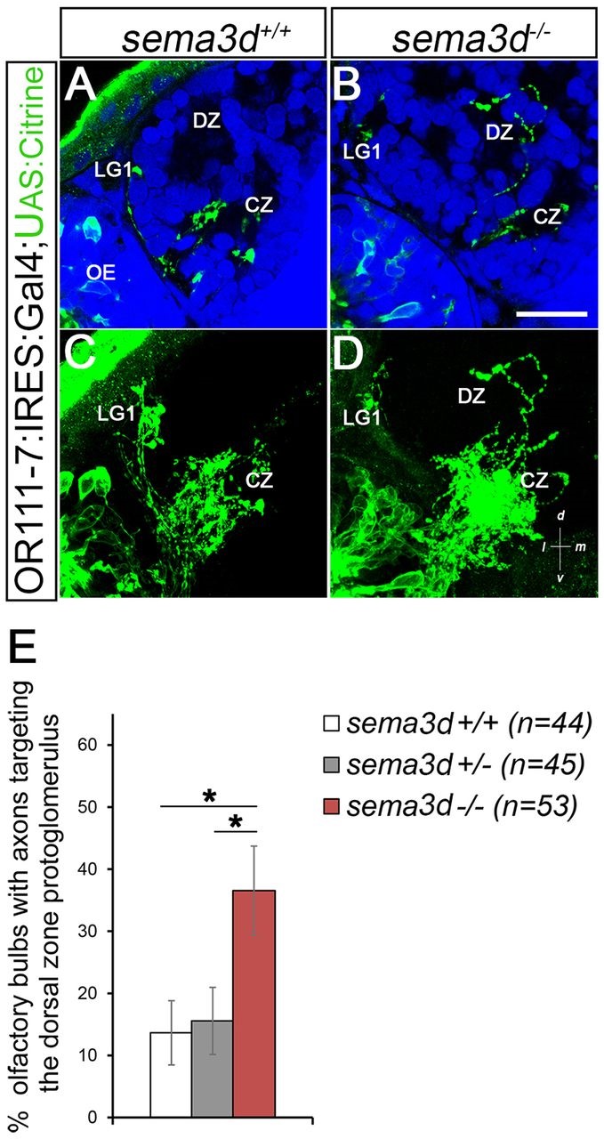
Fig. 2
or111-7 transgenic axons misproject to the DZ in sema3d mutants. (A,B) Single optical sections through 72hpf or111-7:IRES:GAL4;UAS:citrine larvae (frontal view). Axons are in green. Dorsal is up and medial is to the right. Propidium iodide (blue) labels cell bodies, revealing protoglomeruli as cell-free regions. (C,D) Maximum intensity projections of serial optical sections from the larvae shown in A,B. (A,C) Wild-type or111-7 transgenic axons project to the CZ and LG1. (B,D) In sema3d mutants, a subset of or111-7 transgenic axons inappropriately projects to the DZ. Scale bar: 25µm. (E) The percentage of OBs that have a labeled projection to the DZ protoglomerulus. sema3d homozygous mutants are compared with heterozygous and wild-type siblings. Statistical significance was estimated using two-tailed Fisher′s exact test (P<0.05*). Error bars represent s.e. of the sample proportion. CZ, central zone; DZ, dorsal zone; LG1, lateral protoglomerulus 1; OE, olfactory epithelium.