
|
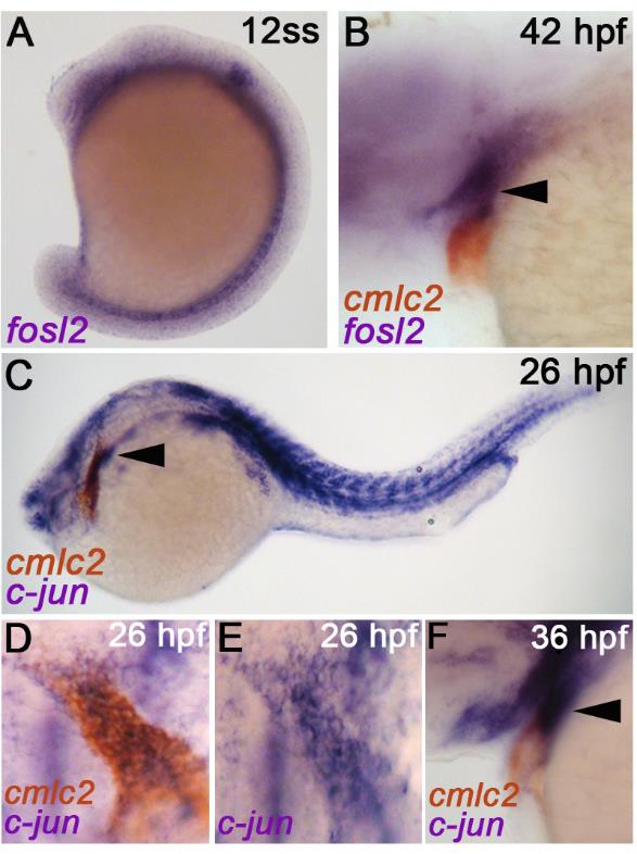
Fig. S4
Developmental expression profiles of AP-1 components fosl2, c-jun, and c-fos.
(A, B) In situ hybridization analysis of fosl2 transcripts at the 12 somites stage (ss) (A) and 42 hours post fertilization (hpf) (B). The embryo shown in (B) is co-stained for the myocardial transcript cmlc2. The arrowhead in (B) highlights fosl2 expression in pharyngeal mesoderm on the extra-cardiac side of the arterial pole where SHF progenitors reside. (C-F) Double in situ hybridization analysis of c-jun and cmlc2 transcripts at 26 hpf (C-E) and 36 hpf (F). Double stained embryos shown in (D) were washed with methanol to remove the cmlc2 signal and photographed again (E). C-jun+ cells were observed on either side of the arterial pole. Arrowhead in (F) highlights c-jun expression in pharyngeal mesoderm on the extra cardiac side of the arterial pole where SHF progenitors reside. In all cases, greater than 20 embryos were analyzed.