
IMAGE
Fig. 4
- ID
- ZDB-IMAGE-160215-11
- Publication
- Edmunds et al., 2016 - Phenoscape: Identifying candidate genes for evolutionary phenotypes
- All Figures
- Figures for Edmunds et al., 2016
Image
|
Figure Caption
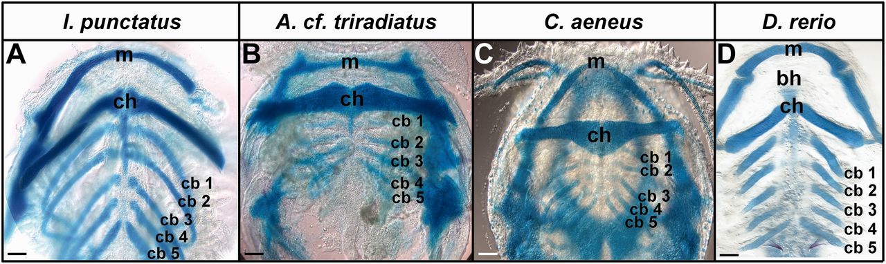
Fig. 4
Pharyngeal arches develop later in catfish than in zebrafish. Chondrification of jaw (mandibular arch), hyoid arch, and branchial arches as detected by Alcian Blue staining. Images show lower branchial elements in ventral view. (A) Ictalurus punctatus, 96 hpf; (B) Ancistrus cf. triradiatus, 96 hpf; (C) Corydoras aeneus, 102 hpf; (D) Danio rerio, 72 hpf. m, Meckel’s cartilage; bh, basihyal; ch, ceratohyal cartilage; cb, ceratobranchial cartilages. Scale bars = 100 µm.
Acknowledgments
This image is the copyrighted work of the attributed author or publisher, and
ZFIN has permission only to display this image to its users.
Additional permissions should be obtained from the applicable author or publisher of the image.
Full text @ Mol Bio Evol