
Image
|
Figure Caption
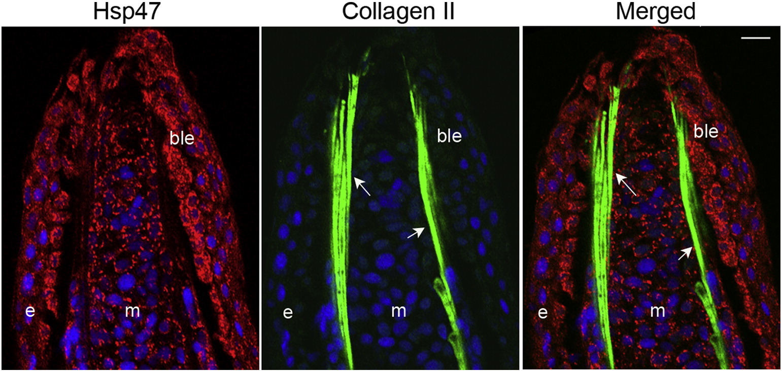
Fig. 9
Localization of Hsp47 and Collagen type II in WT-5 dpa fins. Confocal images of a WT-5 dpa longitudinal fin section immunostained with Hsp47 (red) and collagen type II (green) and counterstained with To-pro to detect nuclei (blue). Arrows indicate actinotrichia. ‘ble’ is basal layer of epithelium, ‘e’ epidermis and ‘m’ mesenchyme. Scale bar is 10µm and applies to all panels.
Figure Data
Acknowledgments
This image is the copyrighted work of the attributed author or publisher, and
ZFIN has permission only to display this image to its users.
Additional permissions should be obtained from the applicable author or publisher of the image.
Reprinted from Mechanisms of Development, 138 Pt 3, Bhadra, J., Iovine, M.K., Hsp47 mediates Cx43-dependent skeletal growth and patterning in the regenerating fin, 364-74, Copyright (2015) with permission from Elsevier. Full text @ Mech. Dev.