
|
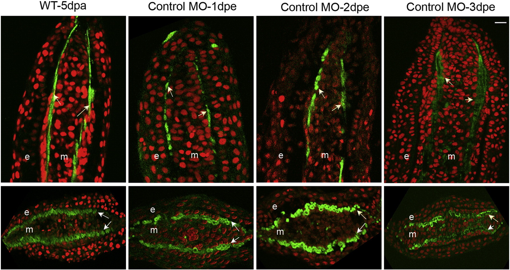
Fig. 10
Examination of actinotrichia in fin sections following injection with standard control MO at different timepoints. Single plane confocal sections were obtained for longitudinal and transverse sections immunostained with Collagen type II (green) and counterstained with propidium iodide (red). Untreated fins are labeled as ‘WT-5 dpa’. Longitudinal (top) and transverse (bottom) sections show regeneration of actinotrichia at different time points following injection and electroporation with standard control MO. Arrows indicate actinotrichia, ‘e’ is epidermis and ‘m’ mesenchyme. Scale bar is 10µm and applies to all panels.
Reprinted from Mechanisms of Development, 138 Pt 3, Bhadra, J., Iovine, M.K., Hsp47 mediates Cx43-dependent skeletal growth and patterning in the regenerating fin, 364-74, Copyright (2015) with permission from Elsevier. Full text @ Mech. Dev.